Abducens VI
| Description | Labelled | Unlabelled |
|---|---|---|
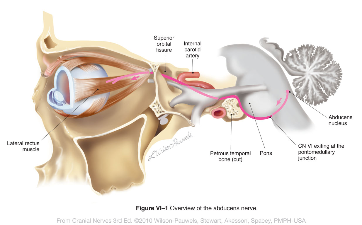
| Figure VI-1 Overview of the abducens nerve. Somatic motor efferent (pink) |
 |
 |
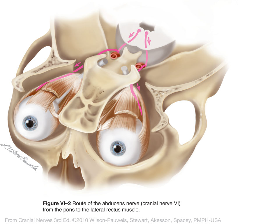
| Figure VI-2 Route of the abducens nerve from the pons to the lateral rectus muscle. Somatic motor efferent (pink) |
 |
 |
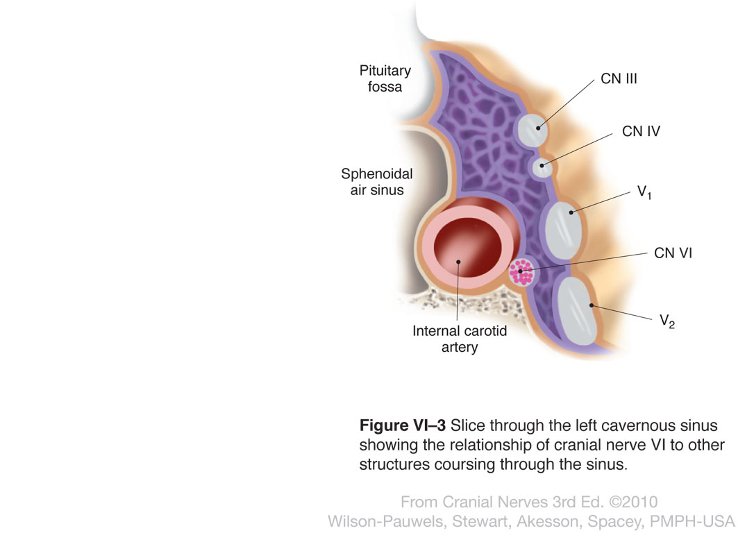
| Figure VI-3 Slice through the left cavernous sinus showing the relationship of cranial nerve VI to other structures coursing through the sinus. Somatic motor efferent (pink) |
 |
 |
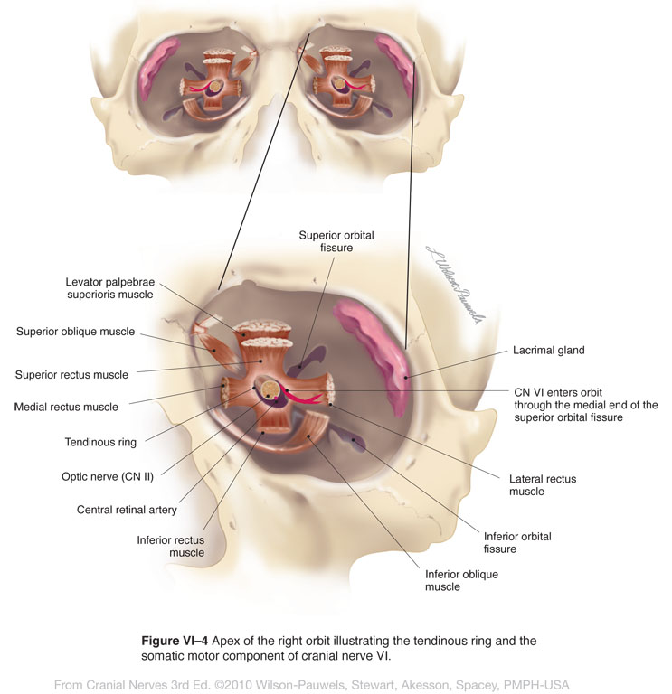
| Figure VI-4 Apex of the right orbit illustrating the tendinous ring and the somatic motor component of CN VI. Somatic motor efferent (pink) |
 |
 |
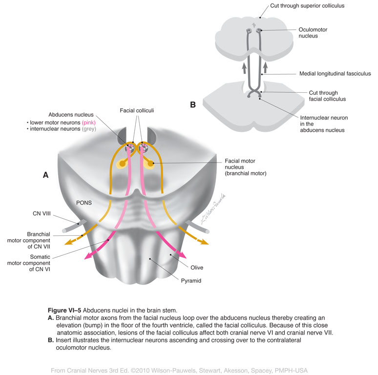
| Figure VI–5 Abducens nuclei in the brain stem. A. Branchial motor axons from the facial nucleus loop over the abducens nucleus thereby creating an elevation (bump) in the floor of the fourth ventricle, called the facial colliculus. Because of this close anatomic association, lesions of the facial colliculus affect both cranial nerve VI and cranial nerve VII. B. Insert illustrates the internuclear neurons ascending and crossing over to the contralateral oculomotor nucleus. Somatic motor efferent (pink) Branchial motor efferent (yellow) |
 |
 |
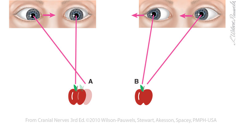
| Figure VI-6 A. On attempted left lateral gaze, Grace was unable to abduct her left eye due to paralysis of her left lateral rectus muscle; therefore, she experienced double vision. B. When looking to the right, Grace was able to direct both eyes toward the same object. |  |
 |
| Figure VI-7 Testing the abducens nerve. |  |
 |