Oculomotor III
| Description | Labelled | Unlabelled |
|---|---|---|
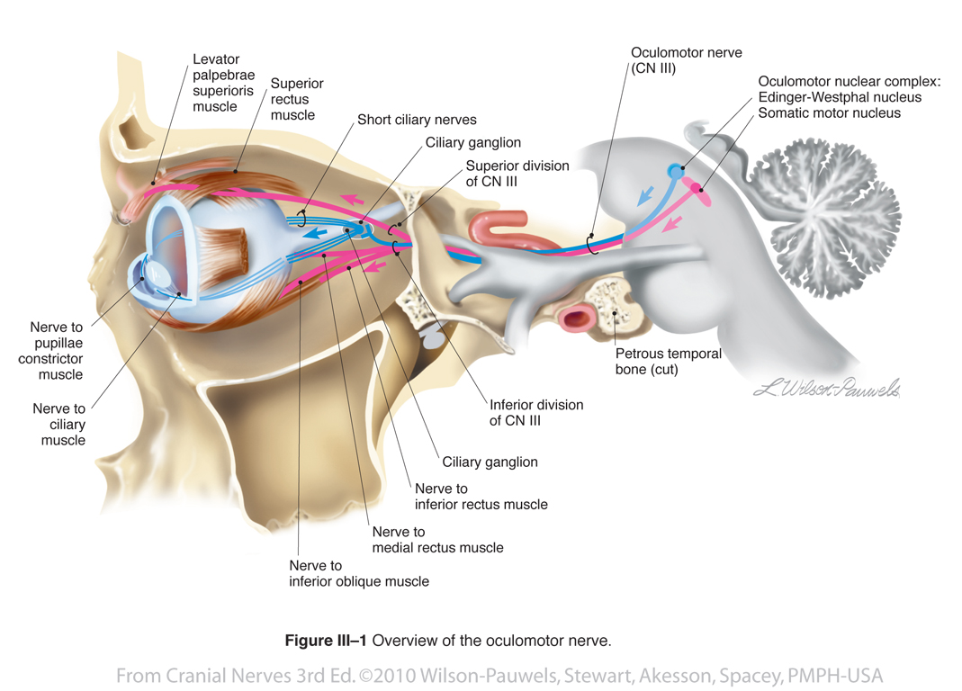
| Figure III-1 Overview of the Oculomotor Nerve. Special sensory afferent (green tracts) to convey visual information from the retina |
 |
 |
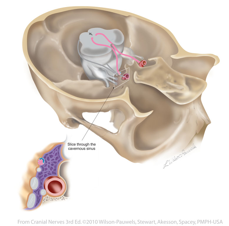
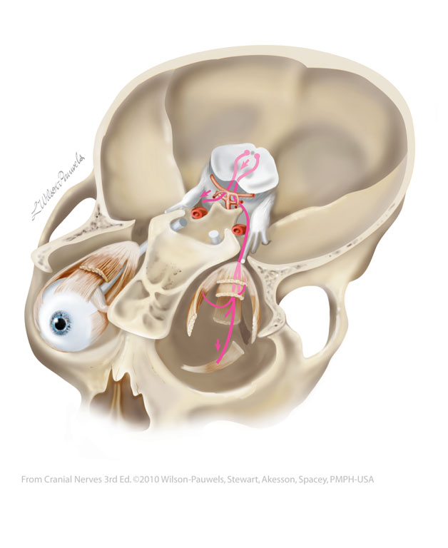
| Figure III-2 Oculomotor nerve coursing through the area of the right cavernous sinus. | 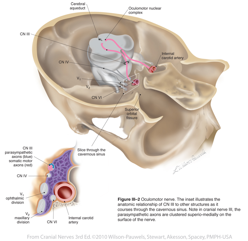 |
 |
| Figure III-3 The somatic motor component of the oculomotor nerve. | 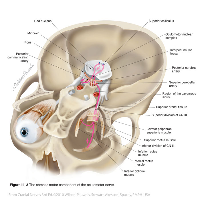 |
 |
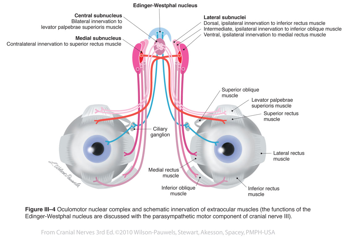
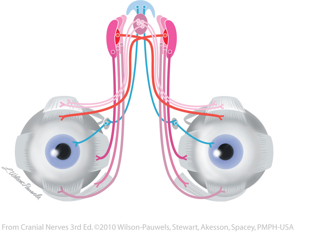
| Figure III-4 Oculomotor nuclear complex and schematic innervation of extraocular muscles. Somatic motor efferent (red tracts) for innervation of muscles of the eye. Visceral motor parasympathetic efferent (blue tracts) to constrictor pupillae and ciliary muscles. |
 |
 |
| Figure III-5 Apex of the right orbit illustrating the tendinous ring | 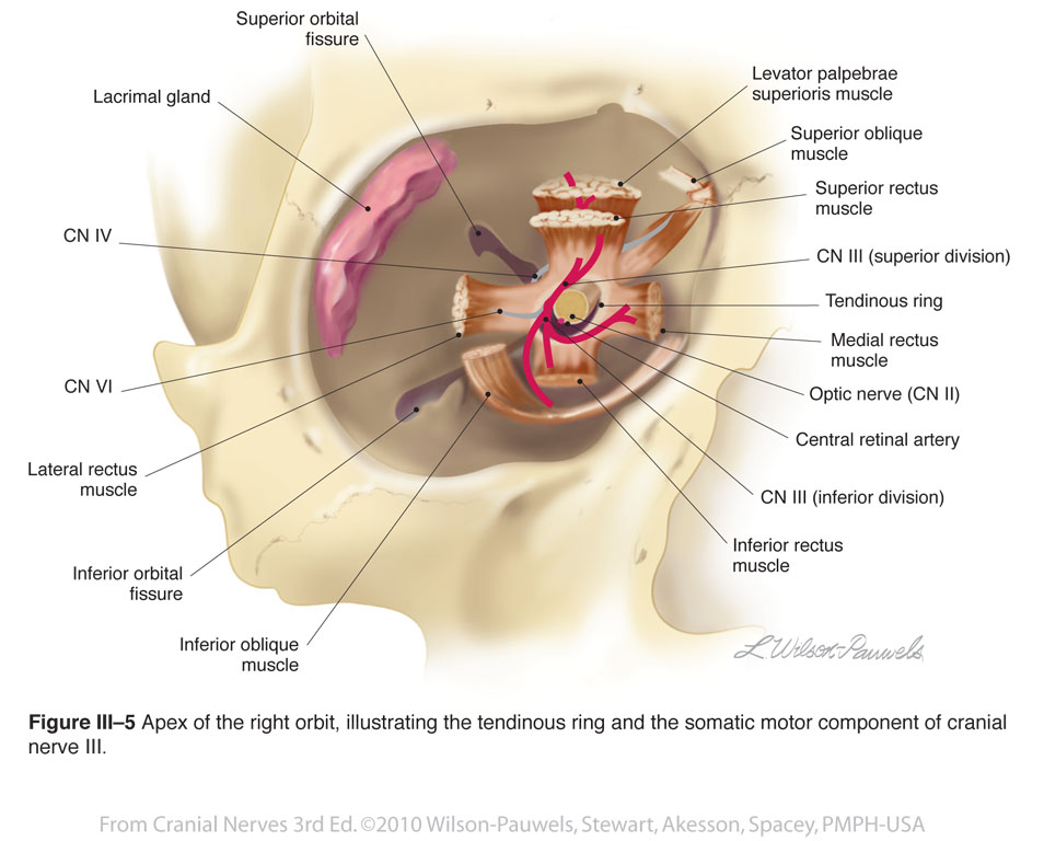 |
 |
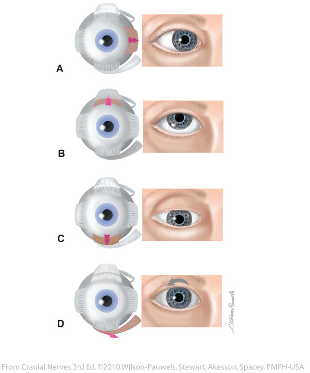
| Figure III-6 Primary actions of the extraocular muscles innervated by cranial nerve III. | 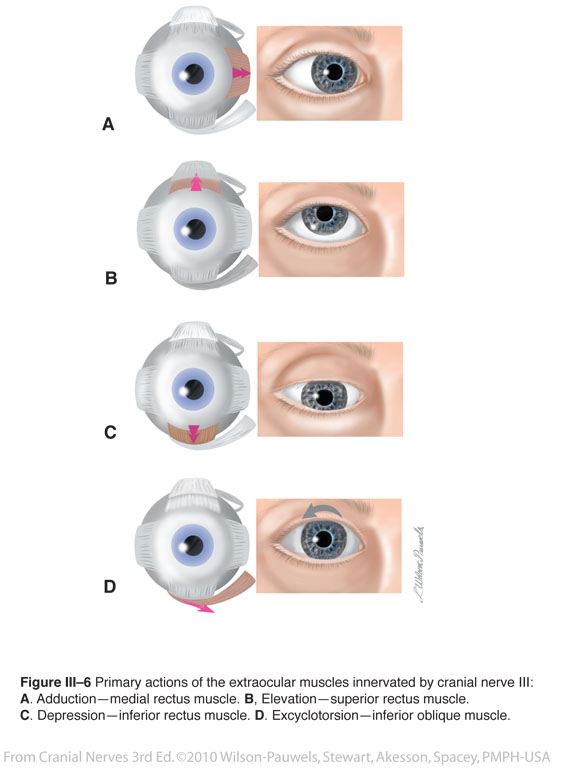 |
 |
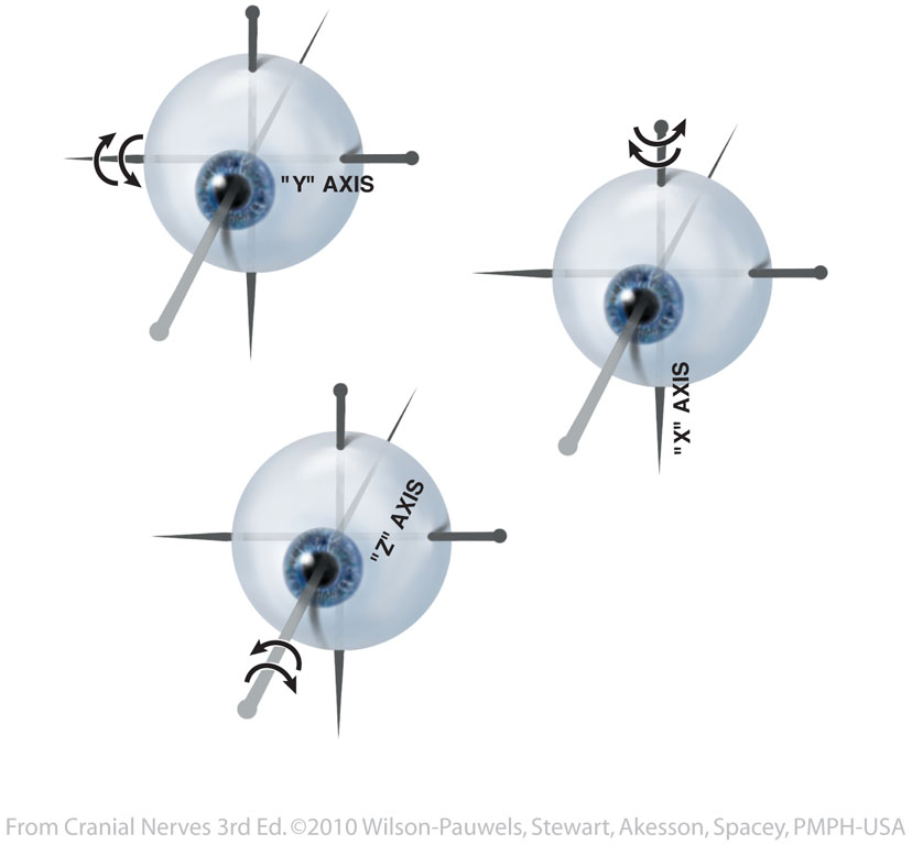
| Figure III-7 Right eye movements around the "X", "Y", and "Z" axes. | 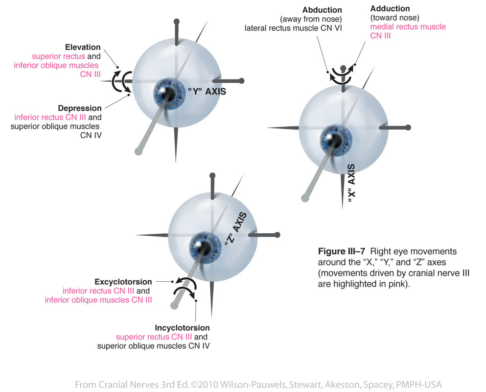 |
 |
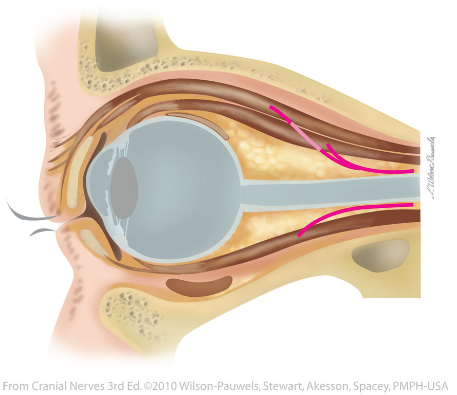
| Figure III-8 Sagittal view of the eye muscles innervated by the oculomotor nerve. |  |
 |
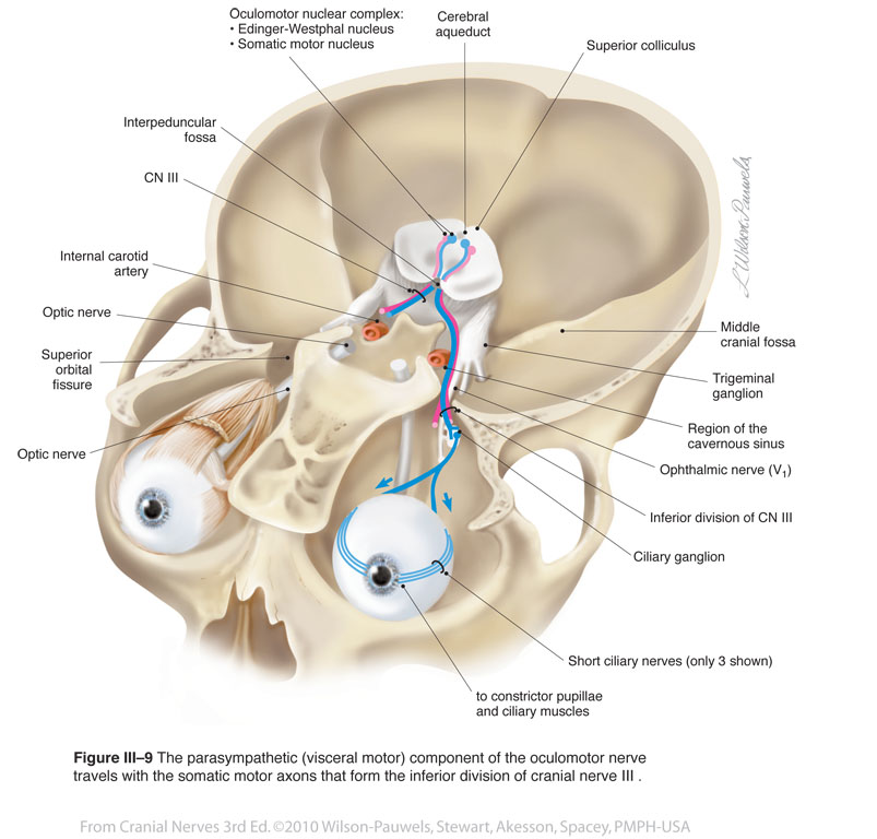
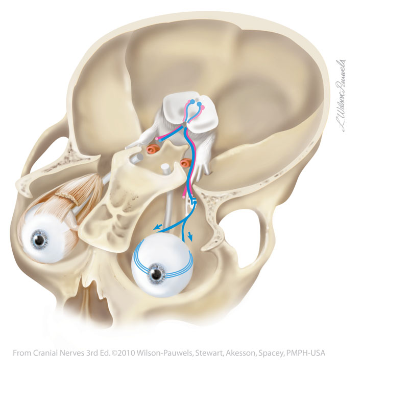
| Figure III-9 The parasympathetic (visceral motor) component of the oculomotor nerve. Somatic motor efferent (red tracts) for innervation of muscles of the eye. Visceral motor parasympathetic efferent (blue tracts) to constrictor pupillae and ciliary muscles. |
 |
 |
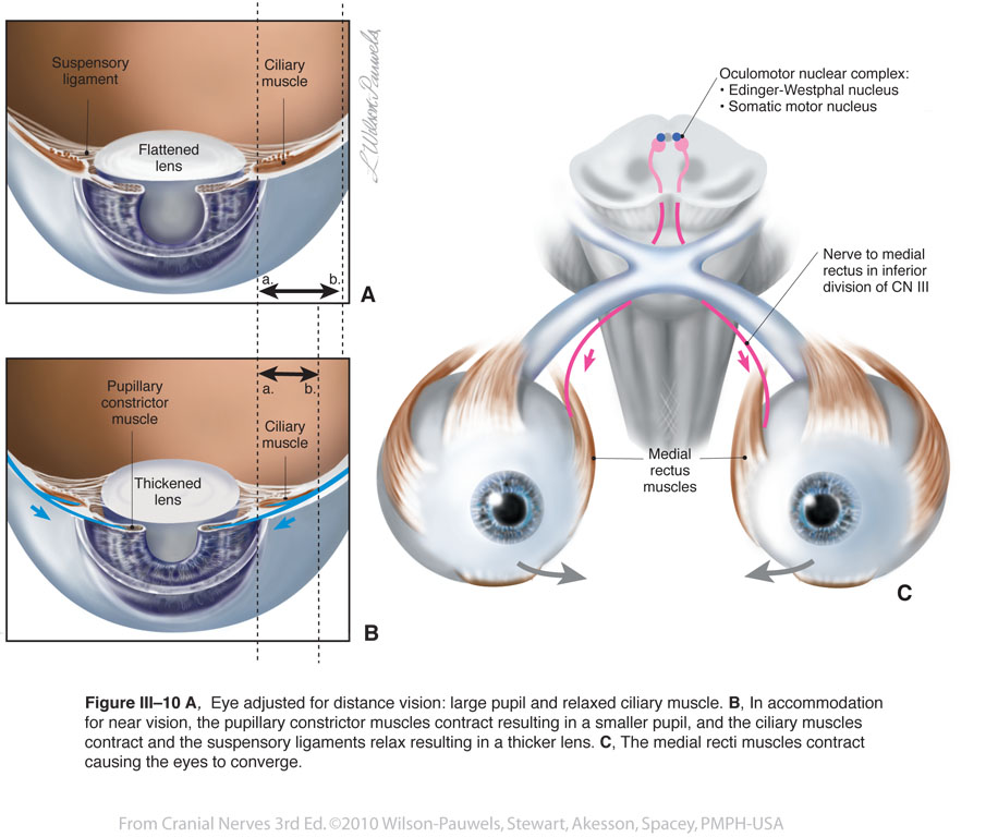
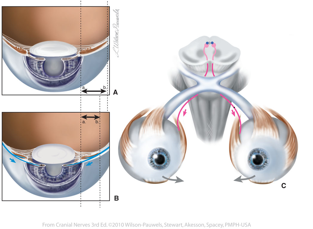
| Figure III-10 Eye adjusted for distance vision & eye adjusted for near vision (accommodation). Somatic motor efferent (red tracts) for innervation of muscles of the eye. Visceral motor parasympathetic efferent (blue tracts) to constrictor pupillae and ciliary muscles. |
 |
 |
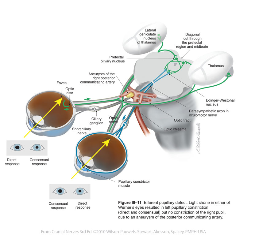
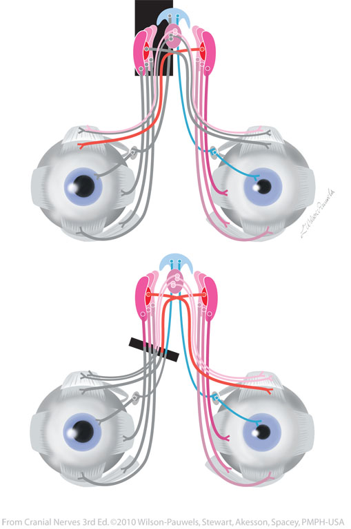
| Figure III-11 Efferent pupillary defect. Somatic motor efferent (red tracts) for innervation of muscles of the eye. Visceral motor parasympathetic efferent (blue tracts) to constrictor pupillae and ciliary muscles. |
 |
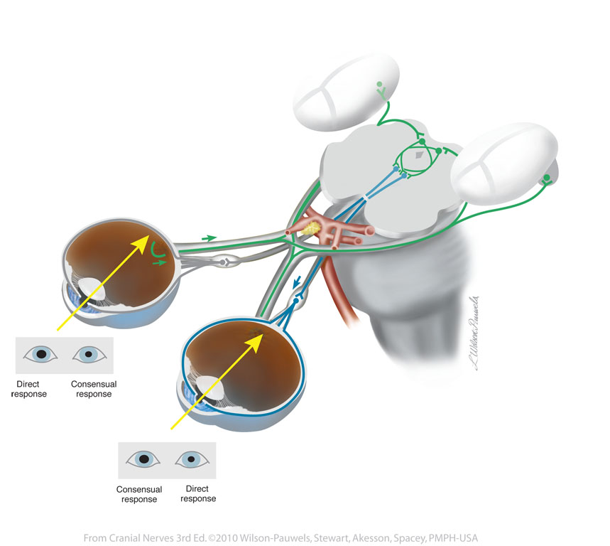 |
| Figure III-12 Relative afferent pupillary defect. Special sensory afferent (green tracts) Parasympathetic motor efferent (blue tracts) |
 |
 |
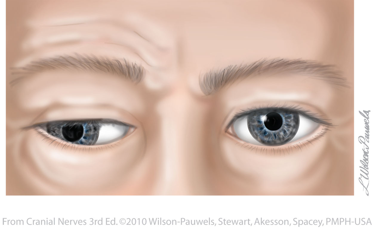
| Figure III-13 Appearance of the eyes in right third nerve palsy. | 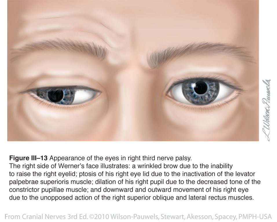 |
 |
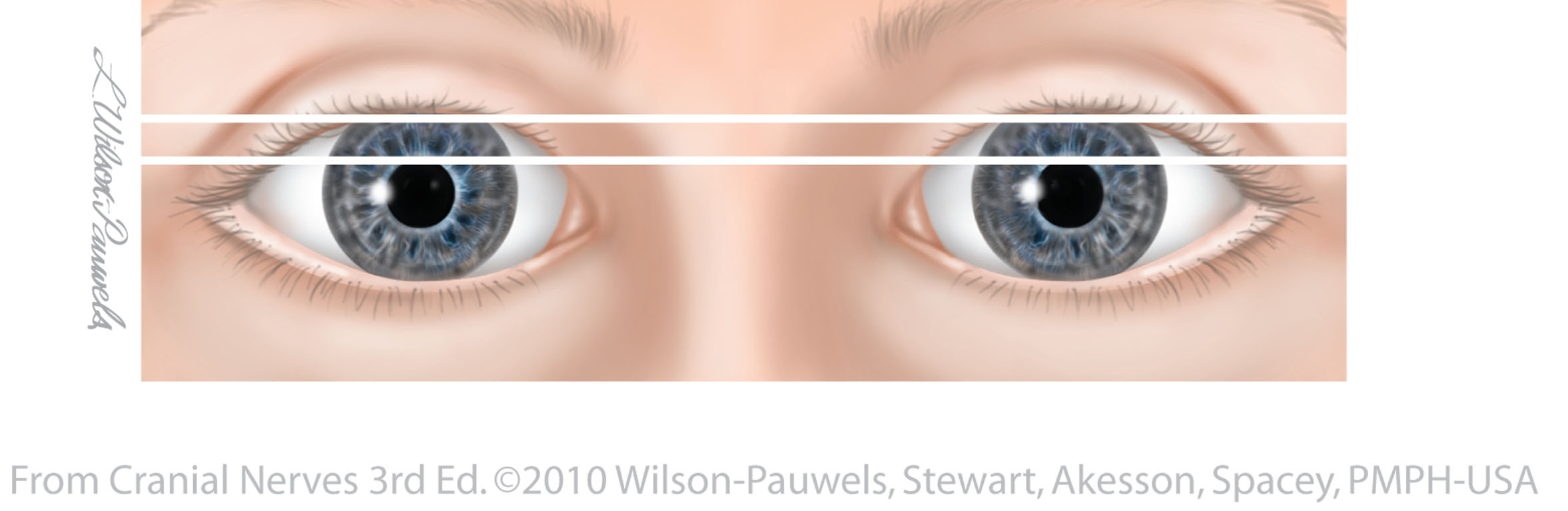
| Figure III-14 Normal eyelid position. |  |
 |
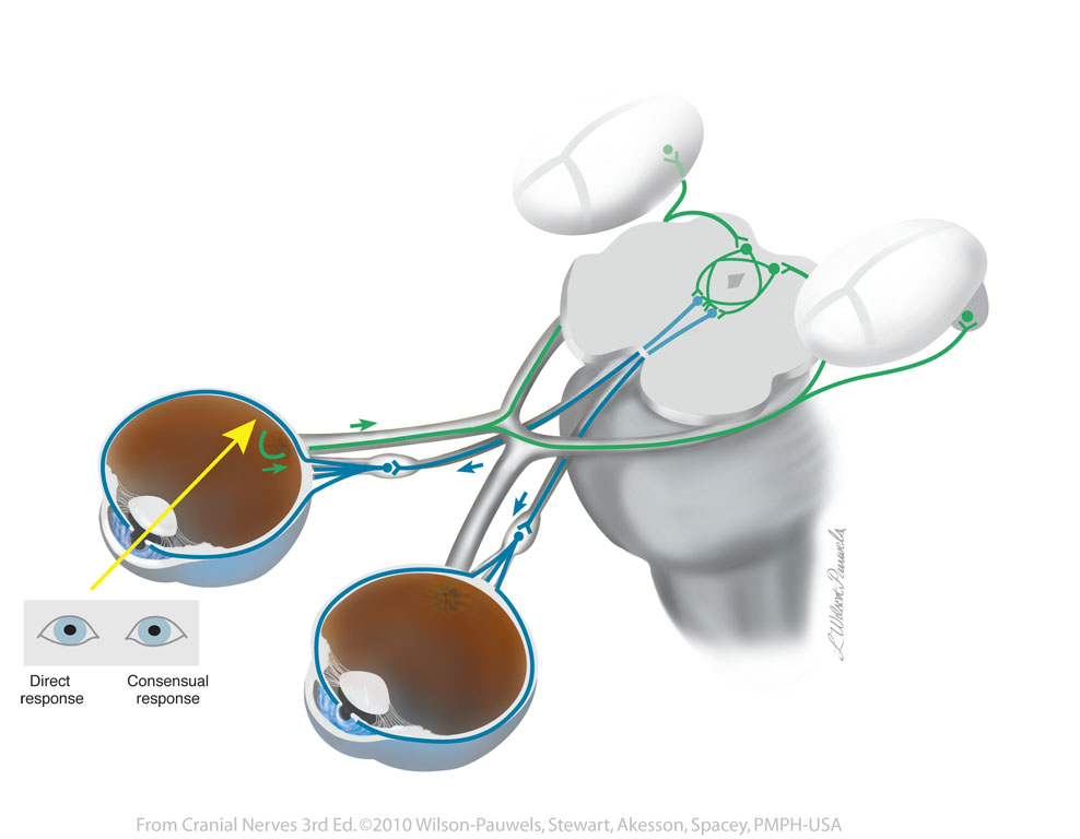
| Figure III-15 Pupillary light reflex. | 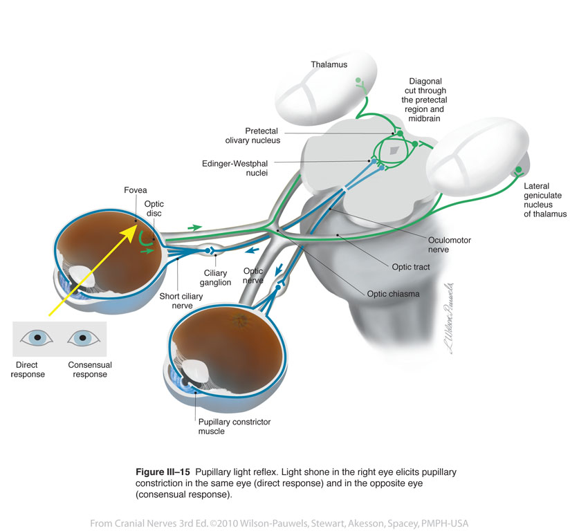 |
 |
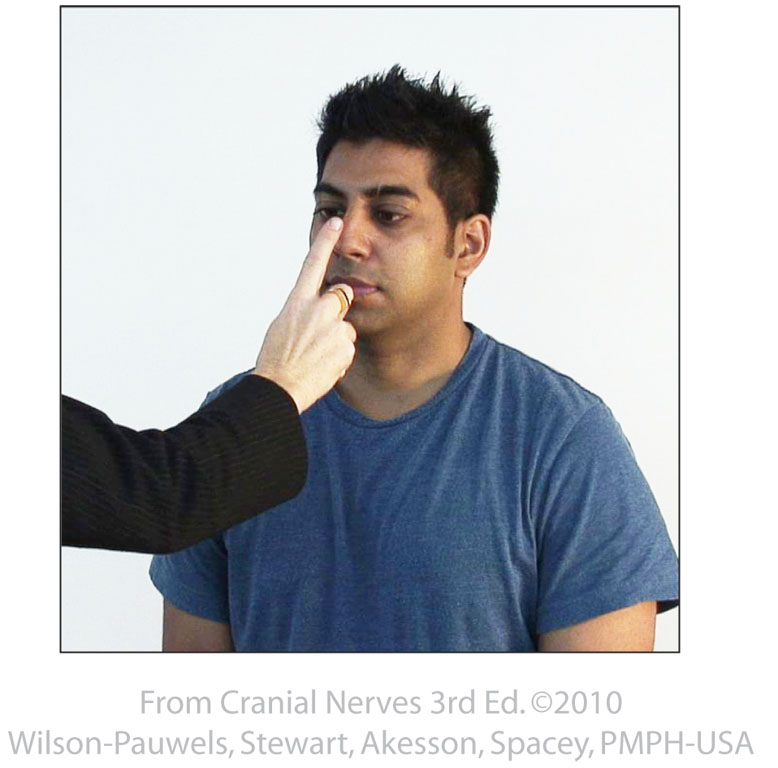
| Figure III-16 Testing cranial nerve III for accommodation. | |
 |