Olfactory I
| Description | Labelled | Unlabelled |
|---|---|---|
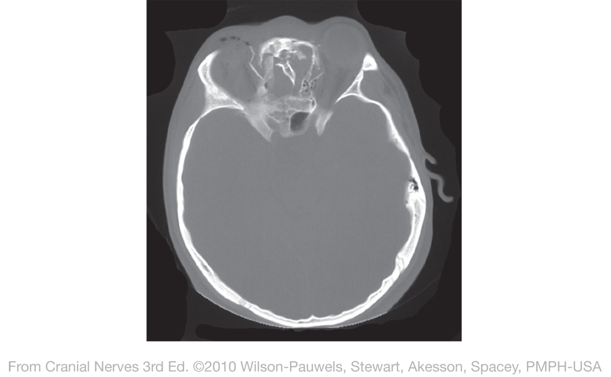
| Figure I-1 Computed tomography (CT) scan through the vomer showing fractures of the vomer, sphenoid, and medial wall of the orbit. (Courtesy of Dr. Robert Nugent, Department of Radiology, Vancouver General Hospital.). | 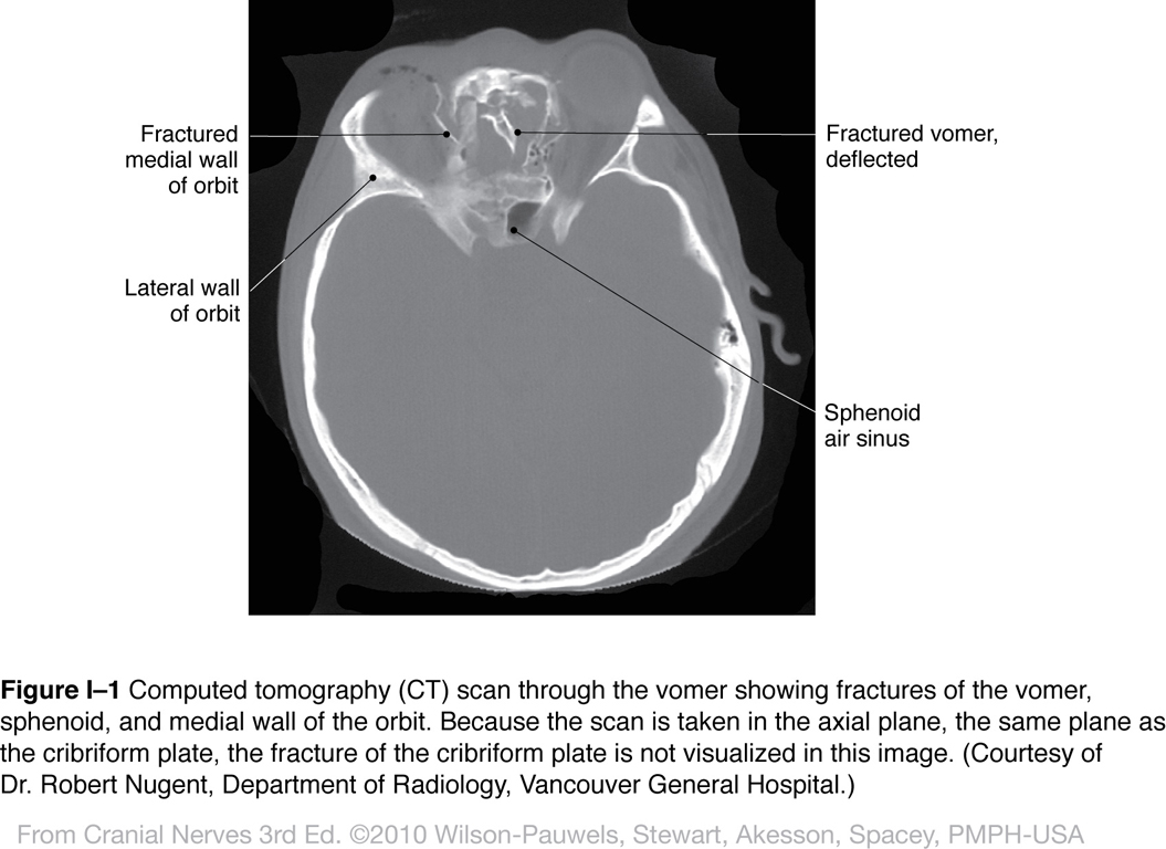 |
 |
| Figure I-2 Olfactory epithelium, bulb, and tracts (structures are enlarged for clarity). Special sensory afferent (green) |
 |
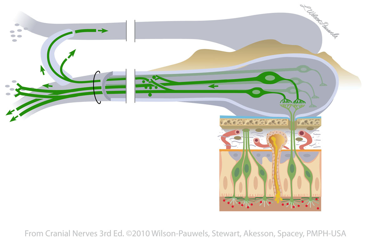 |
| Figure I-3 Schematic olfactory pathway from olfactory epithelium to the olfactory tract. Special sensory afferent (green) |
 |
 |
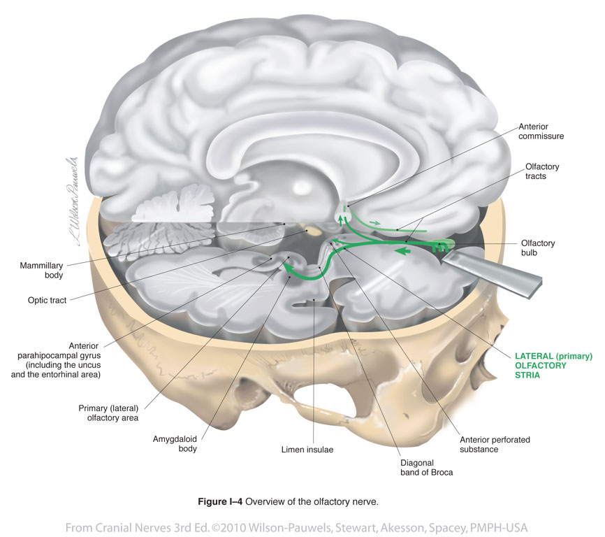
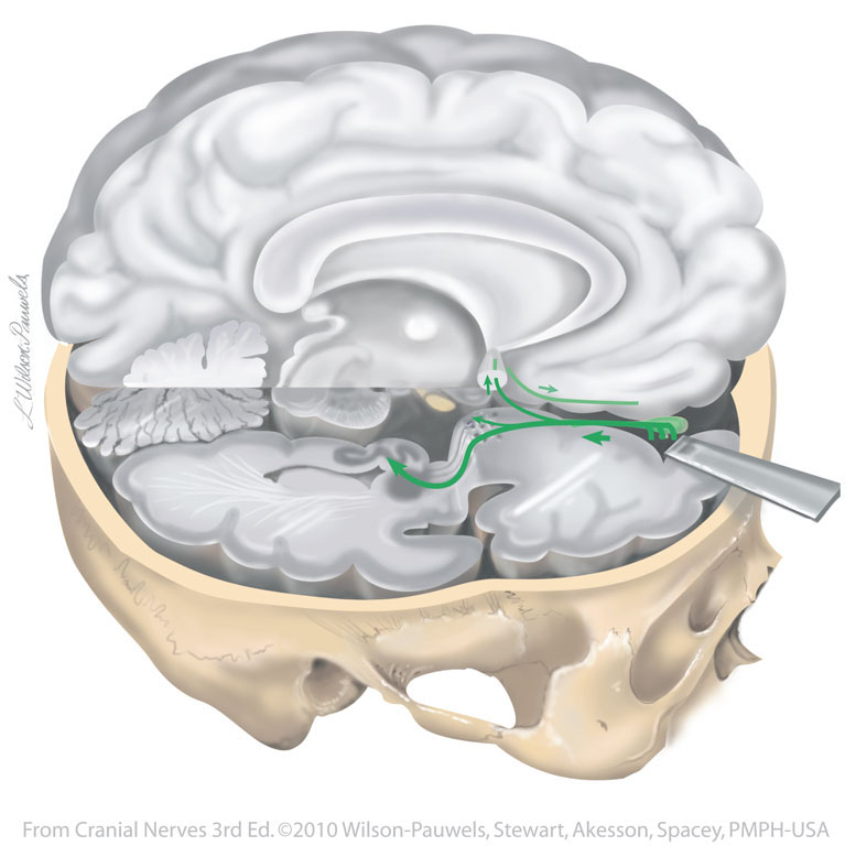
| Figure I-4 Overview of the olfactory nerve. Special sensory afferent (green) |
 |
 |
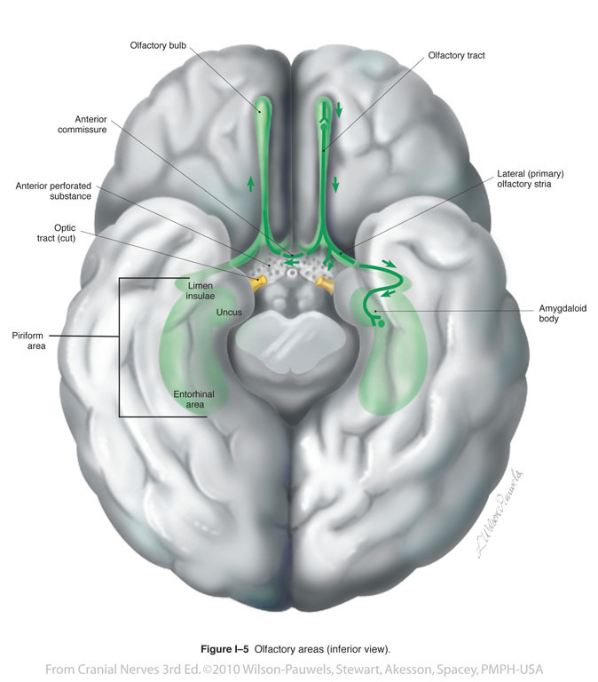
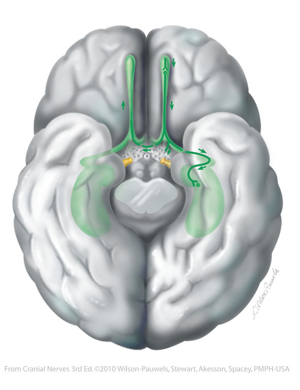
| Figure I-5 Olfactory areas (inferior view). Special sensory afferent (green) |
 |
 |
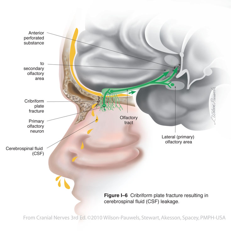
| Figure I-6 Cribriform plate fracture resulting in cerebrospinal fluid (CSF) leakage. Special sensory afferent (green) |
 |
 |
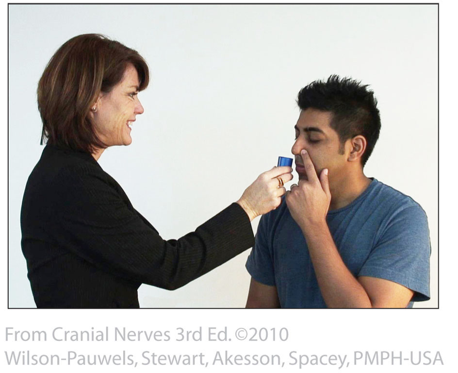
| Figure I-7 Identifying olfactory stimuli to test for anosmia. | 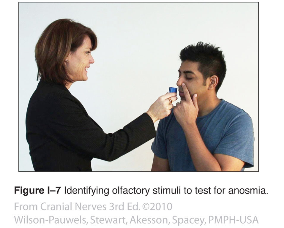 |
 |