| Description |
Labelled |
Unlabelled |
Figure XII–1 Somatic motor component of the hypoglossal nerve (cranial nerve XII).
Somatic motor efferent (pink) |
 |
 |
Figure XII–2 Route of the hypoglossal nerve through the hypoglossal canal to the bifurcation of the internal and external carotid artery.
Somatic motor efferent (pink) |
 |
 |
Figure XII–3 Views of the hypoglossal nucleus. A, Cross-section through the medulla—open portion. B, Ventral view. C, Dorsal view. D, Lateral view.
Somatic motor efferent (pink) |
 |
 |
| Figure XII–4 Carotid artery dissection demonstrating a blood clot between the tunica intima and the tunica media of the internal carotid artery resulting in an enlarged circumference of the artery. |
 |
 |
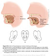
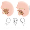
| Figure XII–5 Figure XII–5 Action of the genioglossus muscle in sticking out the tongue. A. Balanced action of both genioglossus muscles is required to stick the tongue out in the midline. B. When the right genioglossus muscle is weak or paralyzed, the left genioglossus muscle pushes the tongue to the weak side. C. When the left genioglossus muscle is weak or paralyzed, the right genioglossus muscle pushes the tongue to the weak side. |
 |
 |
Figure XII–6 CNXII Upper Motor Neuron Lesion (UMNL). Illustration demonstrates contralateral fibers to genioglossus muscles only. In UMNLs, the protruded tongue deviates to the side contralateral to the lesion.
Somatic motor efferent (pink) |
 |
 |
Figure XII–7 CNXII Lower Motor Neuron Lesion (LMNL). Illustration demonstrates contralateral fibers to genioglossus muscles only. In LMNLs, the protruded tongue deviates to the same side as the lesion (this is Todd's lesion).
Somatic motor efferent (pink) |
 |
 |
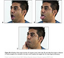
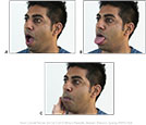
| Figure XII–8 Testing of the tongue involves: A, tongue at rest, frontal view; B, protruding the tongue to observe�if it sticks straight out or deviates to the side; and C, pushing the tongue into the cheek against resistance. |
 |
 |