| Description |
Labelled |
Unlabelled |
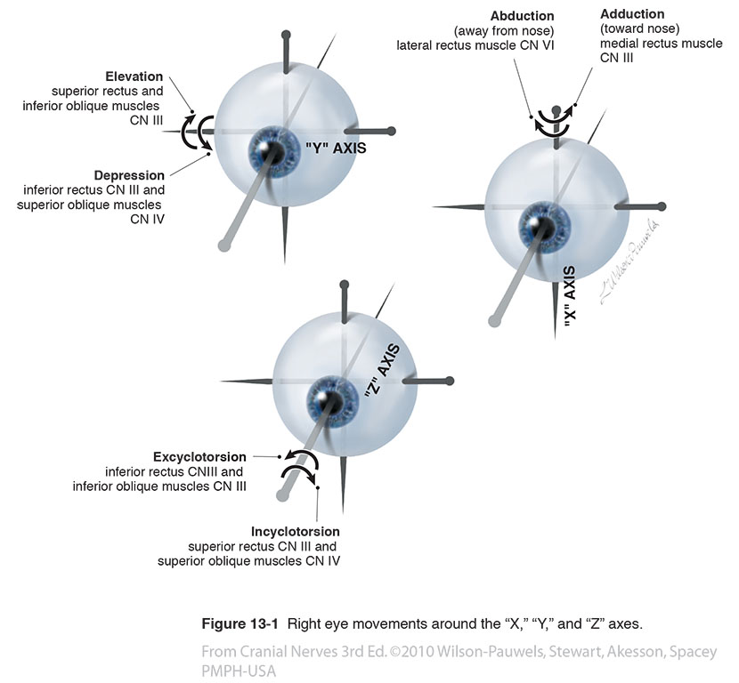
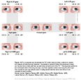
| Figure 13-1 Right eye movements around the “X,” “Y,” and “Z” axes. |
 |
 |
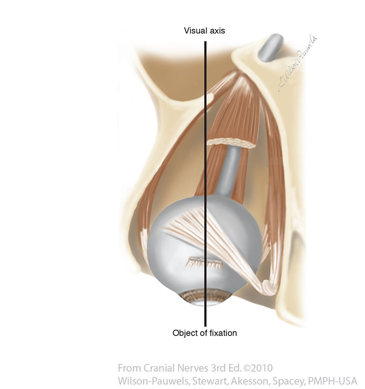
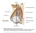
| Figure 13-2 Superior view of the eye and orbit at rest. Superior rectus muscle has been cut to reveal the tendon of the superior oblique muscle. The visual axis, a line connecting the object of fixation and the fovea is indicated. |
 |
 |
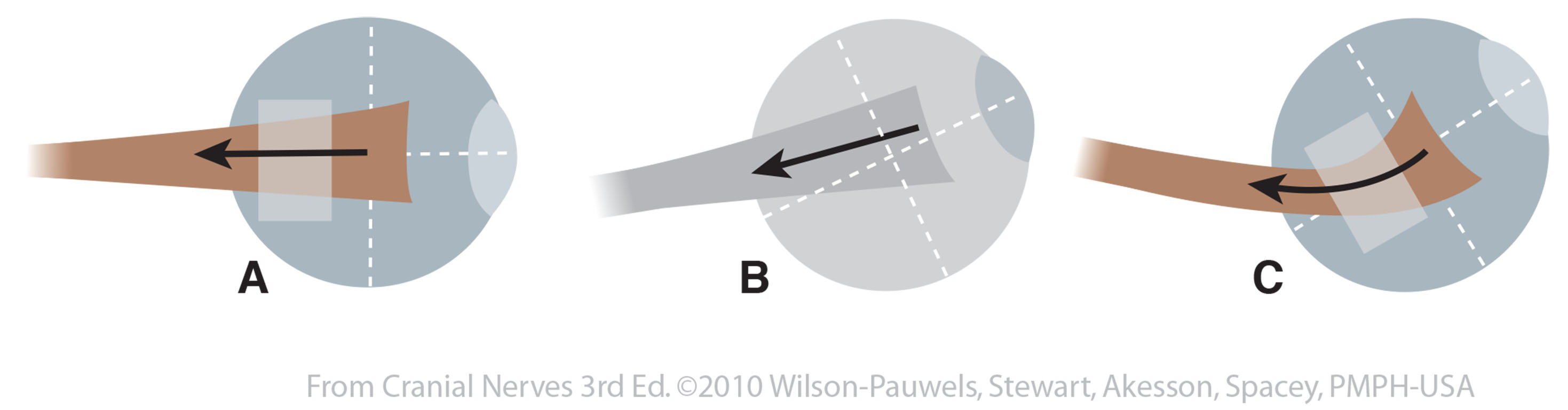
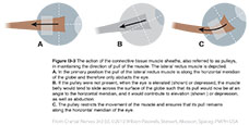
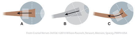
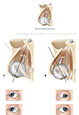
| Figure I3-3 The action of the connective tissue muscle sheaths, also referred to as pulleys, in maintaining the direction of pull of the muscle. |
 |
 |
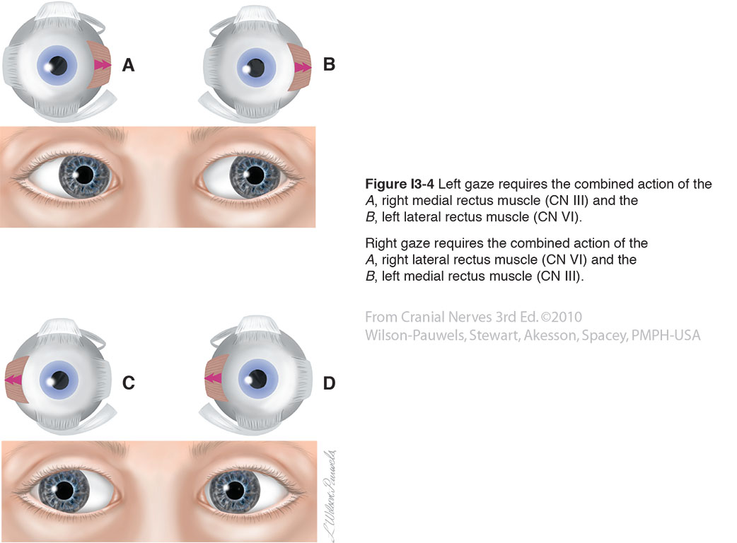
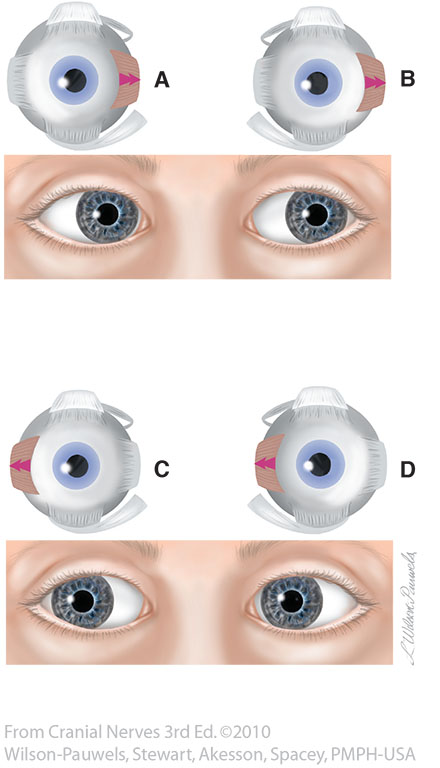
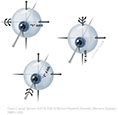
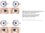
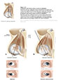
| Figure I3-4 Left gaze requires the combined action of the A, right medial rectus muscle (CN III) and the B, left lateral rectus muscle (CN VI).Right gaze requires the combined action of the A, right lateral rectus muscle (CN VI) and the B, left medial rectus muscle (CN III). |
 |
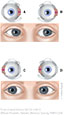 |
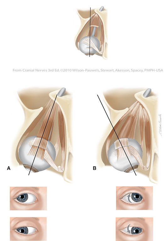
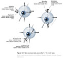
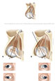
| Figure 13-5 A. Superior and inferior rectus muscles in abduction. B. Superior and inferior rectus muscles in adduction. |
 |
 |
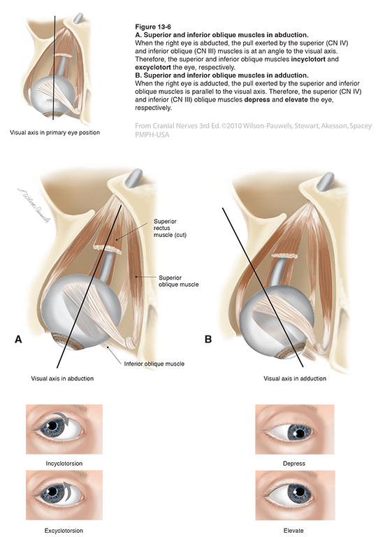
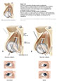
| Figure 13-6 A. Superior and inferior oblique muscles in abduction. B. Superior and inferior oblique muscles in adduction. |
 |
 |
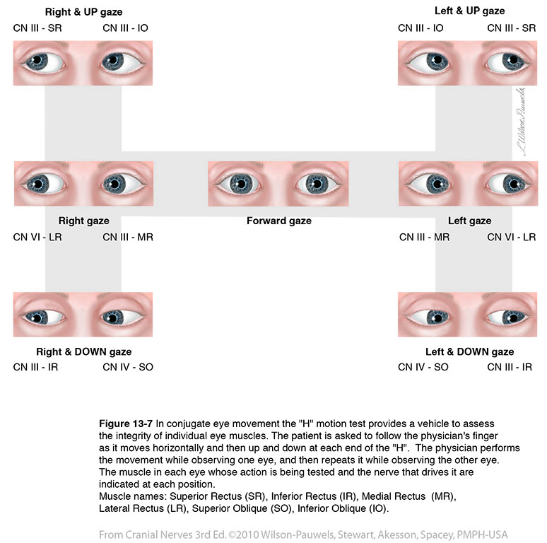
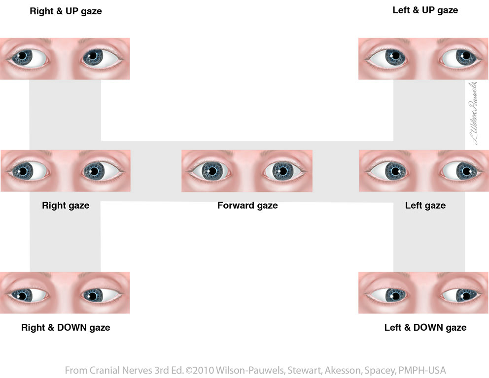
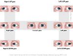
| Figure 13-7 In conjugate eye movement the "H" motion test provides a vehicle to assess the integrity of individual eye muscles. Muscle names: Superior Rectus (SR), Inferior Rectus (IR), Medial Rectus (MR), Lateral Rectus (LR), Superior Oblique (SO), Inferior Oblique (IO). |
 |
 |