Facial VII
| Description | Labelled | Unlabelled |
|---|---|---|
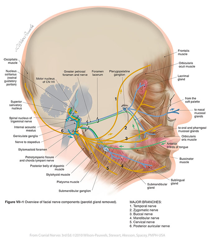
| Figure VII–1 Overview of facial nerve components (parotid gland removed). General sensory afferent (blue-green) Special sensory afferent (green) Branchial motor efferent (yellow) Visceral motor (parasympathetic) efferent (blue) |
 |
 |
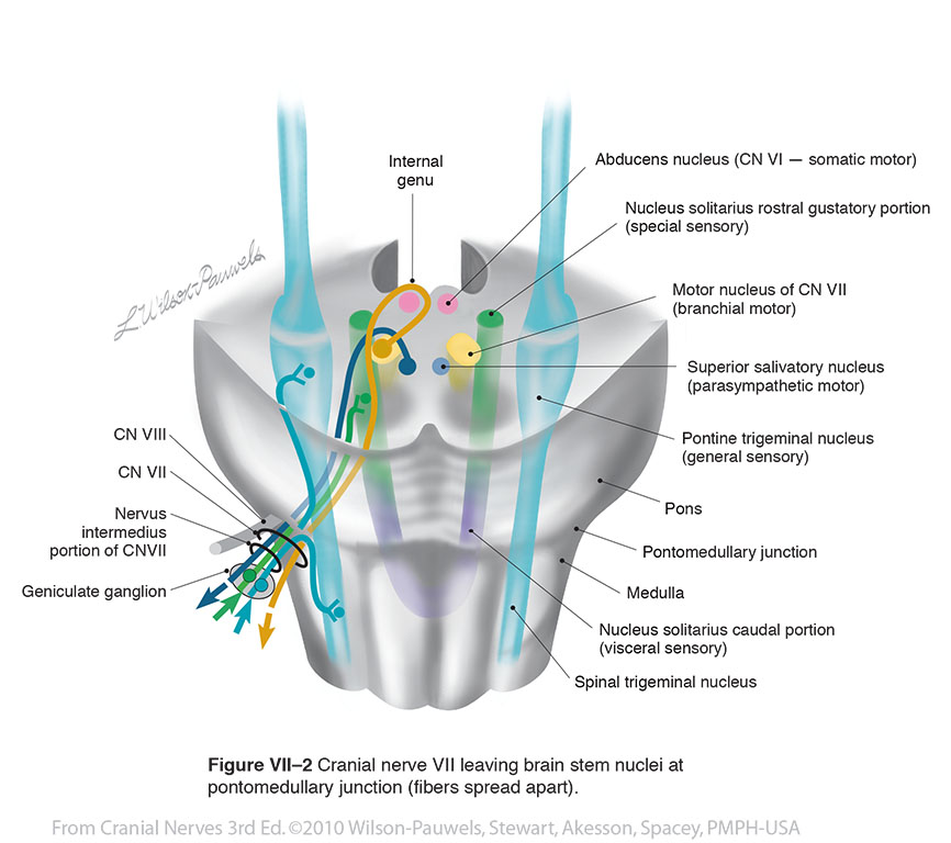
| Figure VII–2 Cranial nerve VII leaving brain stem nuclei at pontomedullary junction (fibers spread apart). General sensory afferent (blue-green) Special sensory afferent (green) Branchial motor efferent (yellow) Visceral motor (parasympathetic) efferent (blue) |
 |
 |
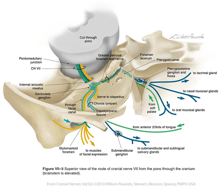
| Figure VII–3 Superior view of the route of cranial nerve VII from the pons through the cranium (brainstem is elevated). General sensory afferent (blue-green) Special sensory afferent (green) Branchial motor efferent (yellow) Visceral motor (parasympathetic) efferent (blue) |
 |
 |
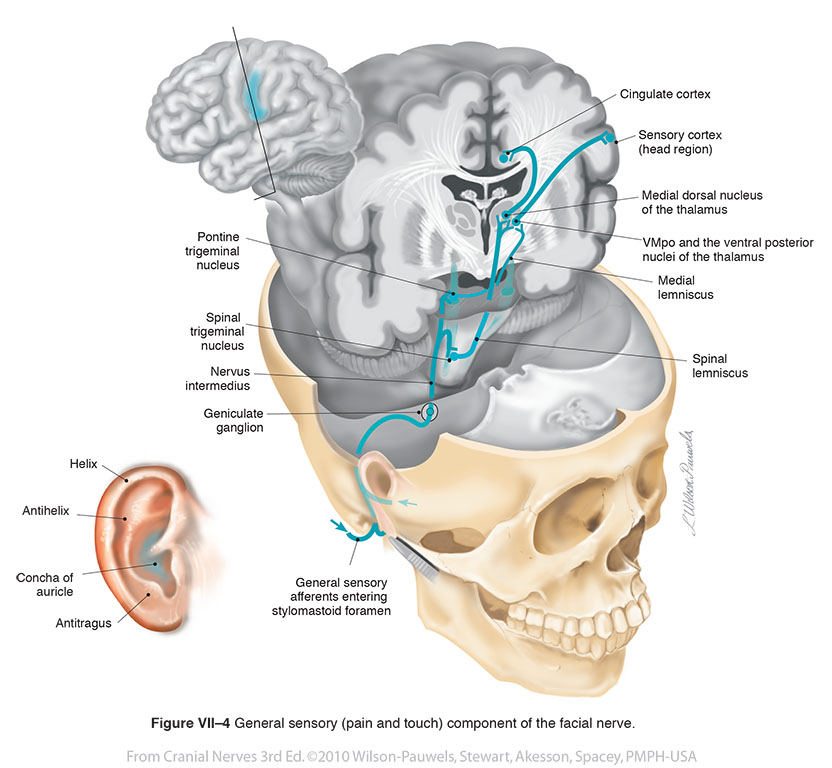
| Figure VII–4 General sensory (pain and touch) component of the facial nerve. General sensory afferent (blue-green) |
 |
 |
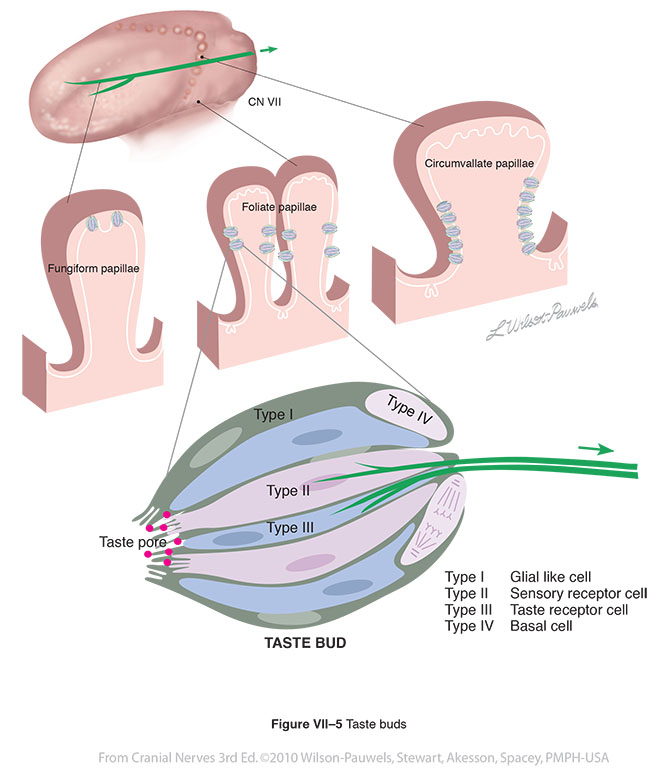
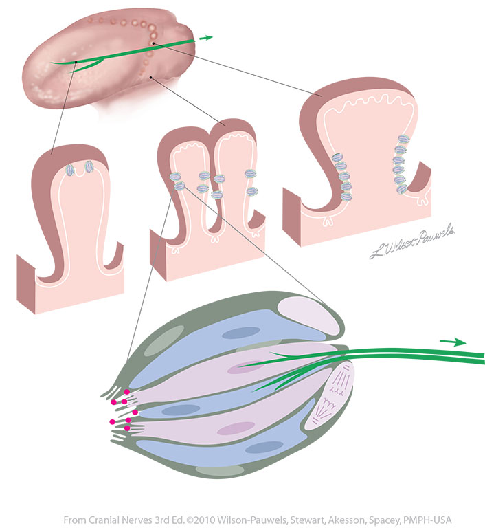
| Figure VII–5 Taste buds. Special sensory afferent (green) |
 |
 |
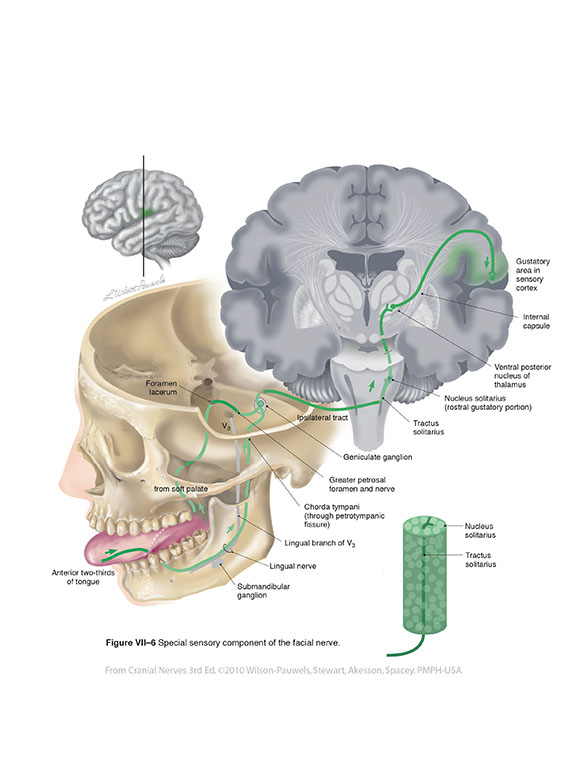
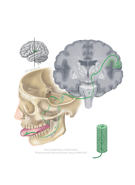
| Figure VII–6 Special sensory component of the facial nerve. Special sensory afferent (green) |
 |
 |
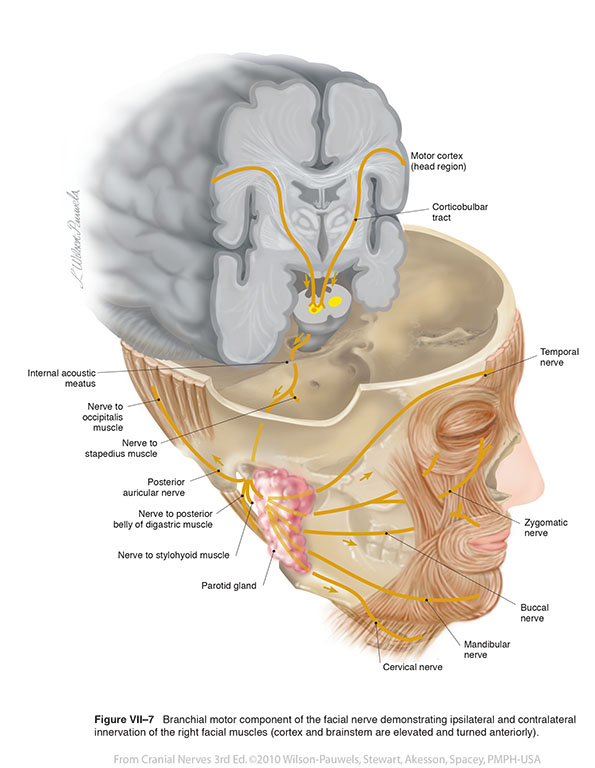
| Figure VII–7 Branchial motor component of the facial nerve demonstrating ipsilateral and contralateral innervation of the right facial muscles (cortex and brainstem are elevated and turned anteriorly). Branchial motor efferent (yellow) |
 |
 |
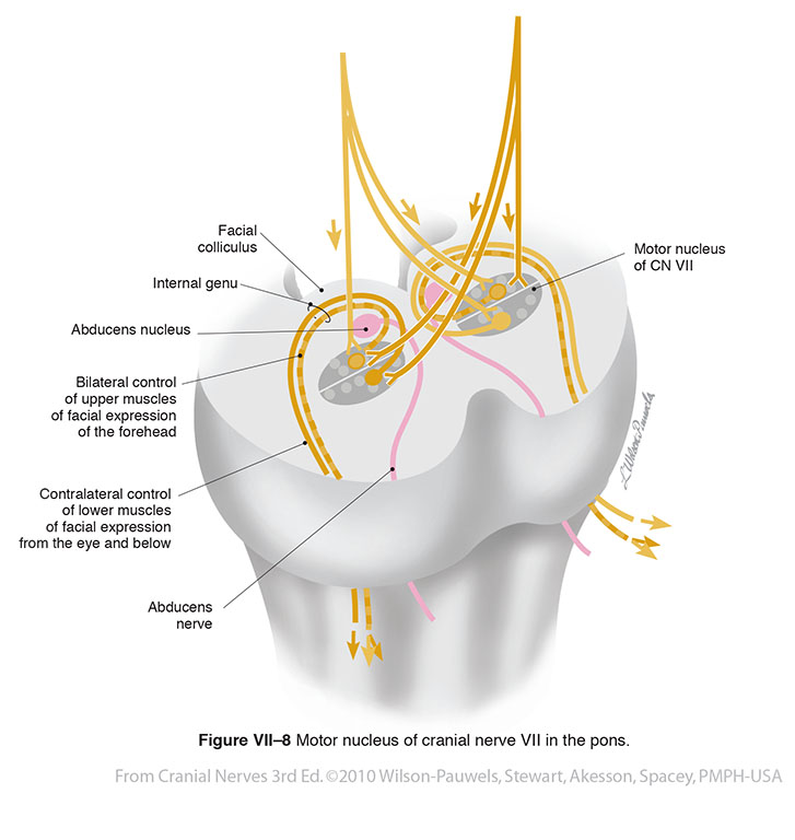
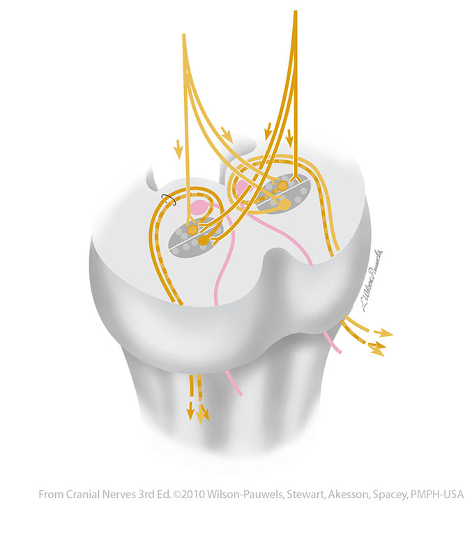
| Figure VII–8 Motor nuclei of cranial nerve VII in the pons. Branchial motor efferent (yellow) |
 |
 |
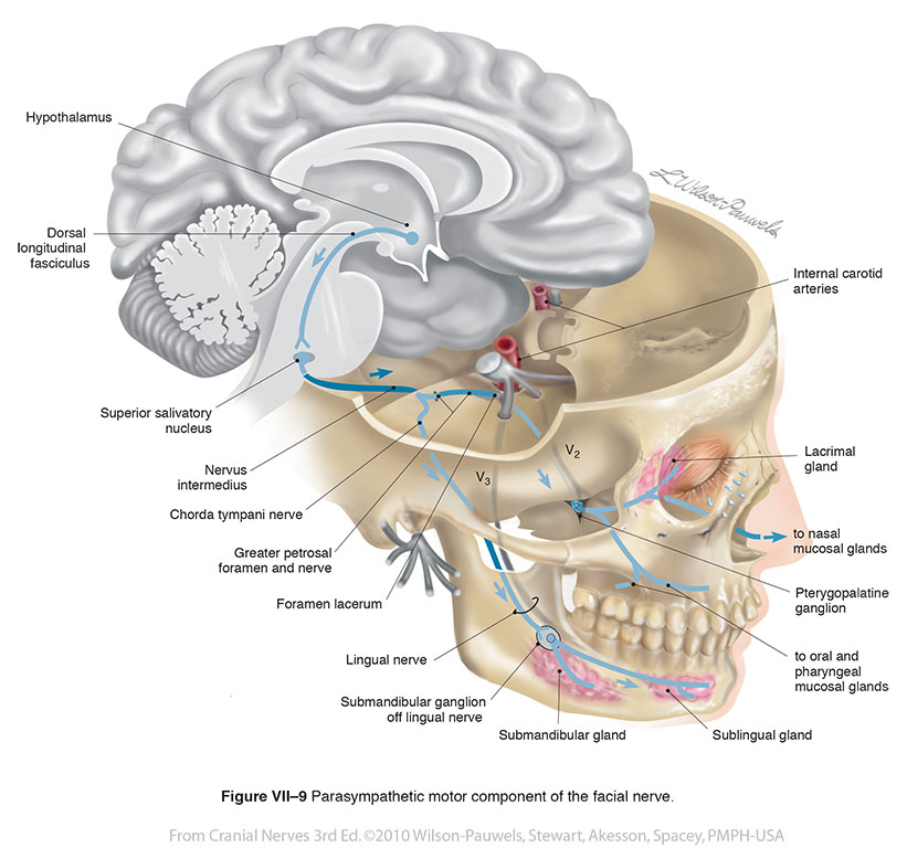
| Figure VII–9 Parasympathetic motor component of the facial nerve. Somatic motor efferent (red tracts) for innervation of muscles of the eye. Visceral motor (parasympathetic) efferent (blue) |
 |
 |
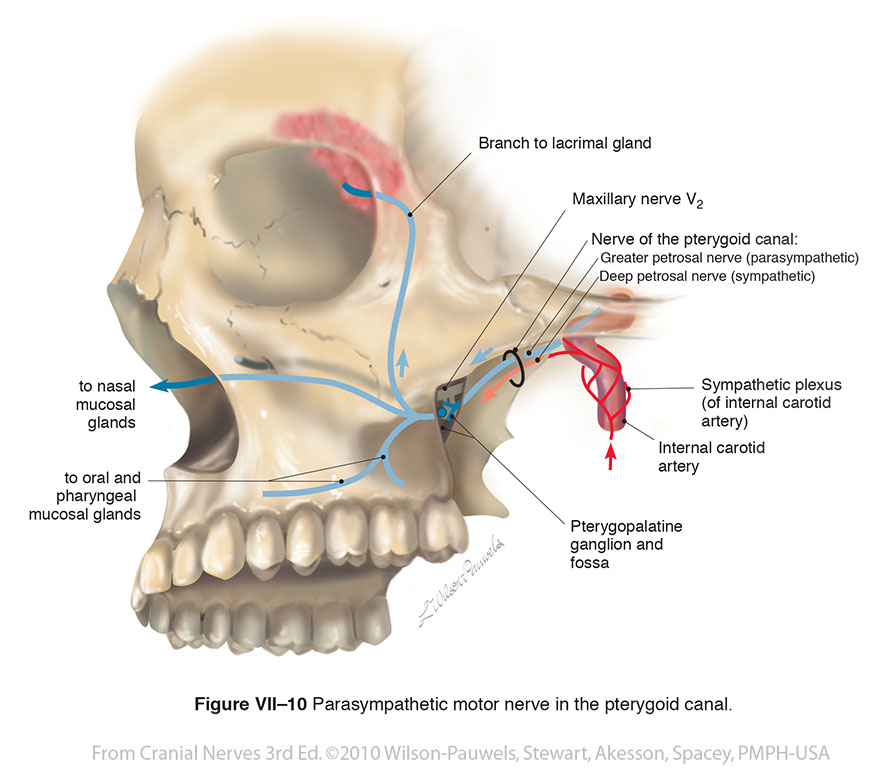
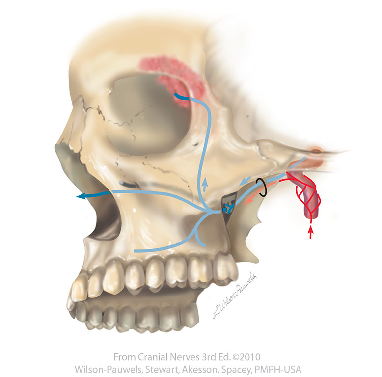
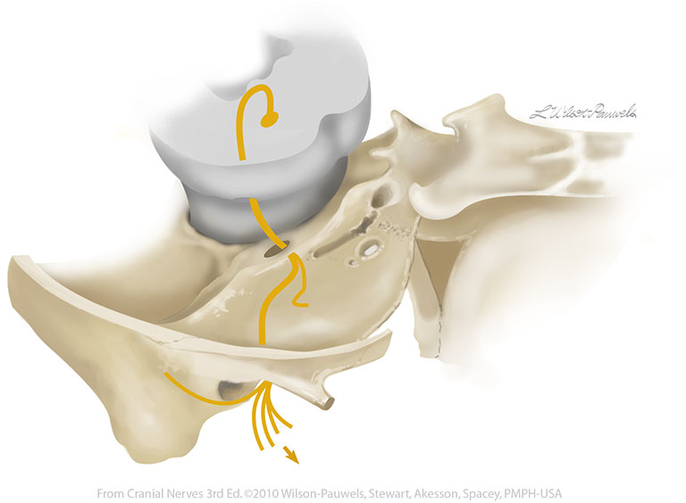
| Figure VII–10 Parasympathetic motor nerve in the pterygoid canal. Visceral motor (parasympathetic) efferent (blue) |
 |
 |
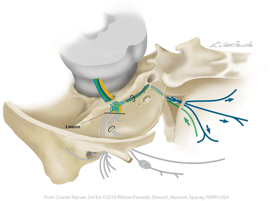
| Figure VII–11 Facial nerve lesion distal to the geniculate ganglion. General sensory afferent (blue-green) Special sensory afferent (green) Branchial motor efferent (yellow) Visceral motor (parasympathetic) efferent (blue) |
 |
 |
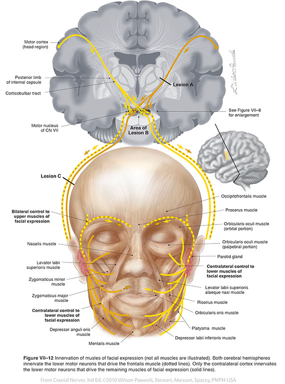
| Figure VII–12 Innervation of musles of facial expression (not all muscles are illustrated). Both cerebral hemispheres innervate the lower motor neurons that drive the frontalis muscle (dotted lines). Only the contralateral cortex innervates the lower motor neurons that drive the remaining muscles of facial expression (solid lines). Branchial motor efferent (yellow) |
 |
 |
| Figure VII–13 Lower Motor Neuron Lesions (LMNLs) affecting the branchial motor axons in A, pons, B, internal acoustic meatus, and C, styloid mastoid foramen (brainstem is elevated) Branchial motor efferent (yellow) |
 |
 |
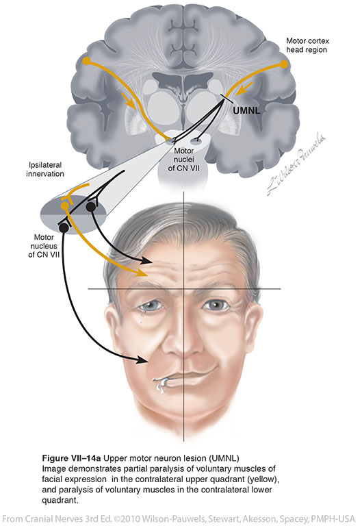
| Figure VII–14a Upper motor neuron lesion (UMNL). Image demonstrates partial paralysis of voluntary muscles of facial expression in the contralateral upper quadrant (yellow), and paralysis of voluntary muscles in the contralateral lower quadrant. Branchial motor efferent (yellow) |
 |
 |
| Figure VII–14b Bilateral and contralateral control from motor nucleus of CNVII. Branchial motor efferent (yellow) |
 |
 |
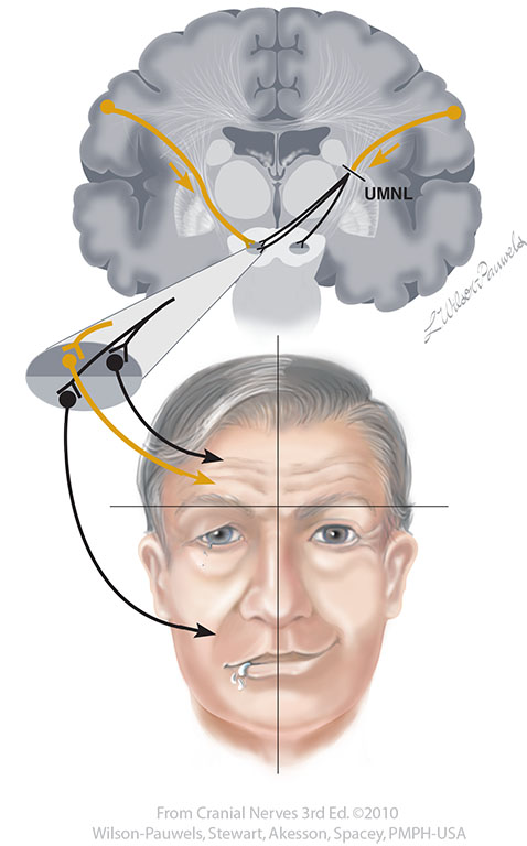
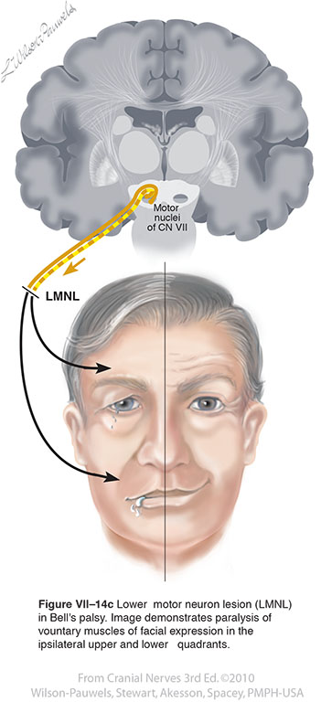
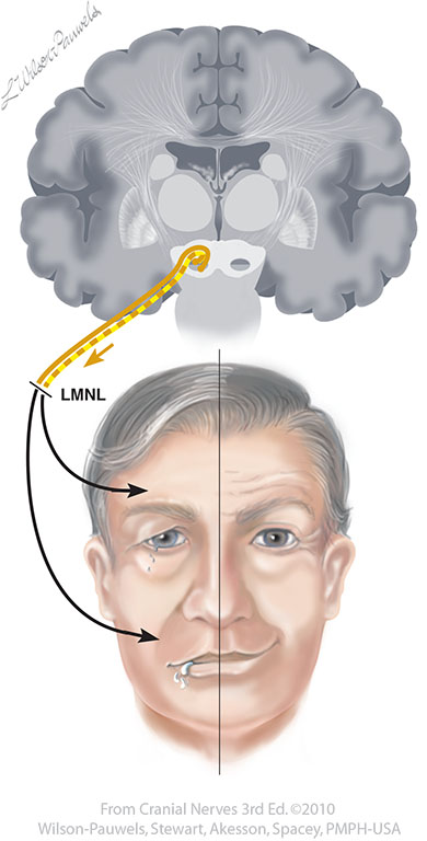
| Figure VII–14c Lower motor neuron lesion (LMNL) in Bell's palsy. Image demonstrates paralysis of vountary muscles of facial expression in the ipsilateral upper and lower quadrants. Branchial motor efferent (yellow) |
 |
 |
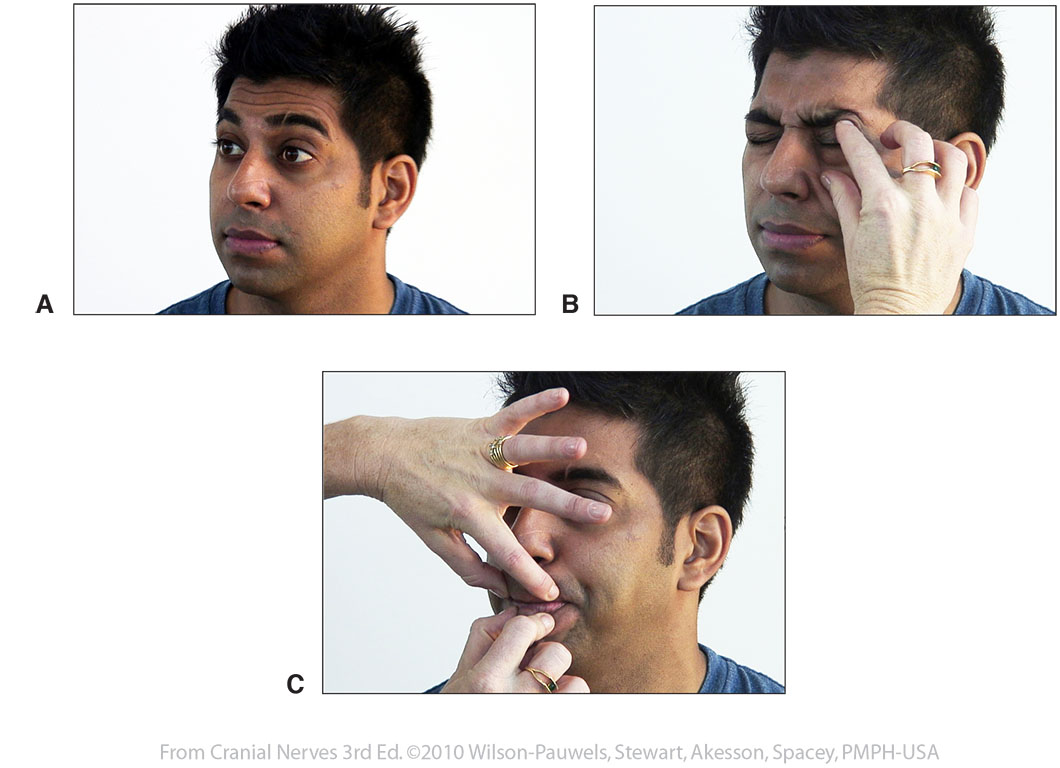
| Figure VII–15 Clinical tests for the muscles of facial expression. A, Frontalis muscle. B, Orbicularis oculi muscle. C, Orbicularis oris muscle. | 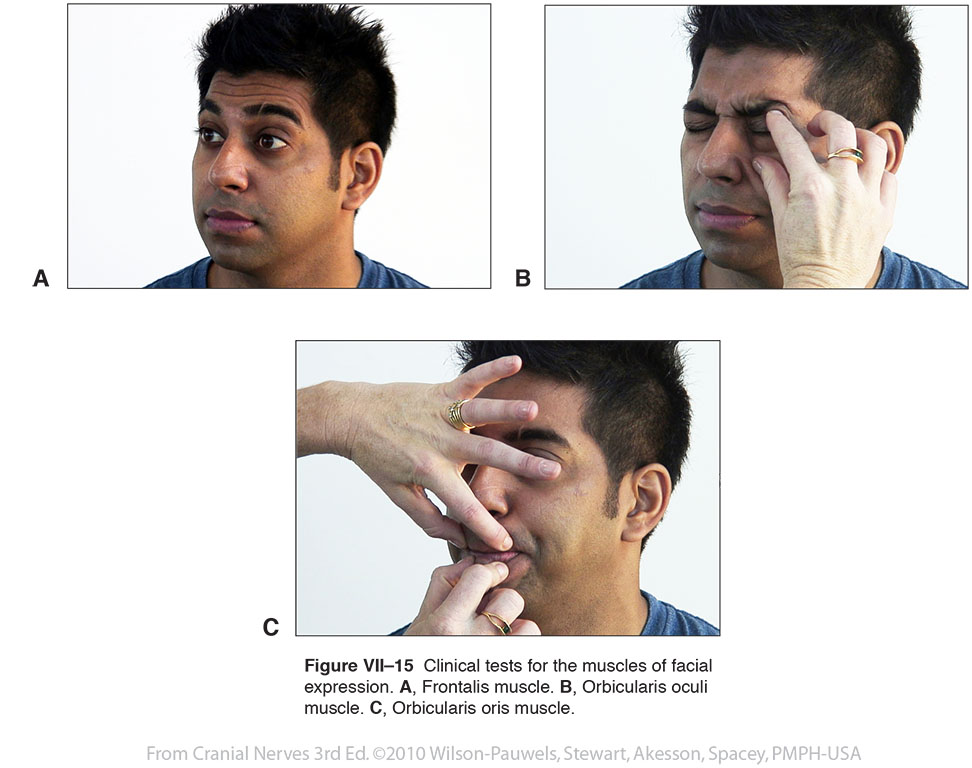 |
 |
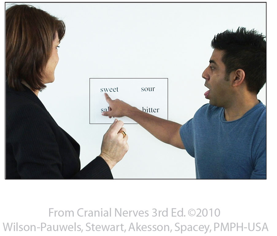
| Figure VII–16 Clinical testing for taste. Four basic modalities (sweet, sour, bitter, and salty) are tested on each side of the tongue. After the examiner holds the tongue and touches one side of it with a cotton swab soaked in a test solution, the patient points to the taste he perceives on the board. | 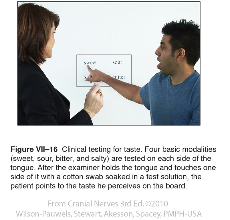 |
 |