Trigeminal V
| Description | Labelled | Unlabelled |
|---|---|---|
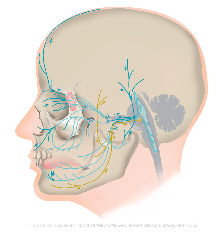
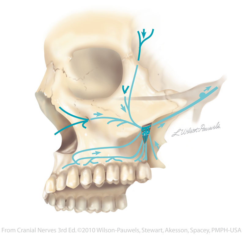
| Figure V-1 Overview of the trigeminal nerve. General sensory afferent (blue-green) Branchial motor efferent (yellow) |
 |
 |
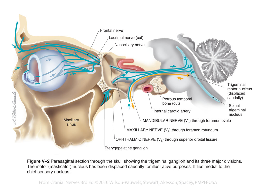
| Figure V–2 Parasagittal section through the skull showing the trigeminal ganglion and its three major divisions. The motor (masticator) nucleus has been displaced caudally for illustrative purposes. It lies medial to the chief sensory nucleus. General sensory afferent (blue-green) |
 |
 |
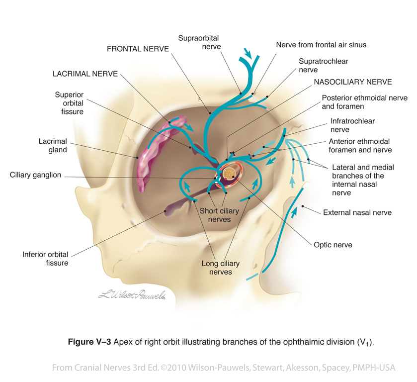
| Figure V–3 Apex of right orbit illustrating branches of the ophthalmic division (V1). General sensory afferent (blue-green) |
 |
 |
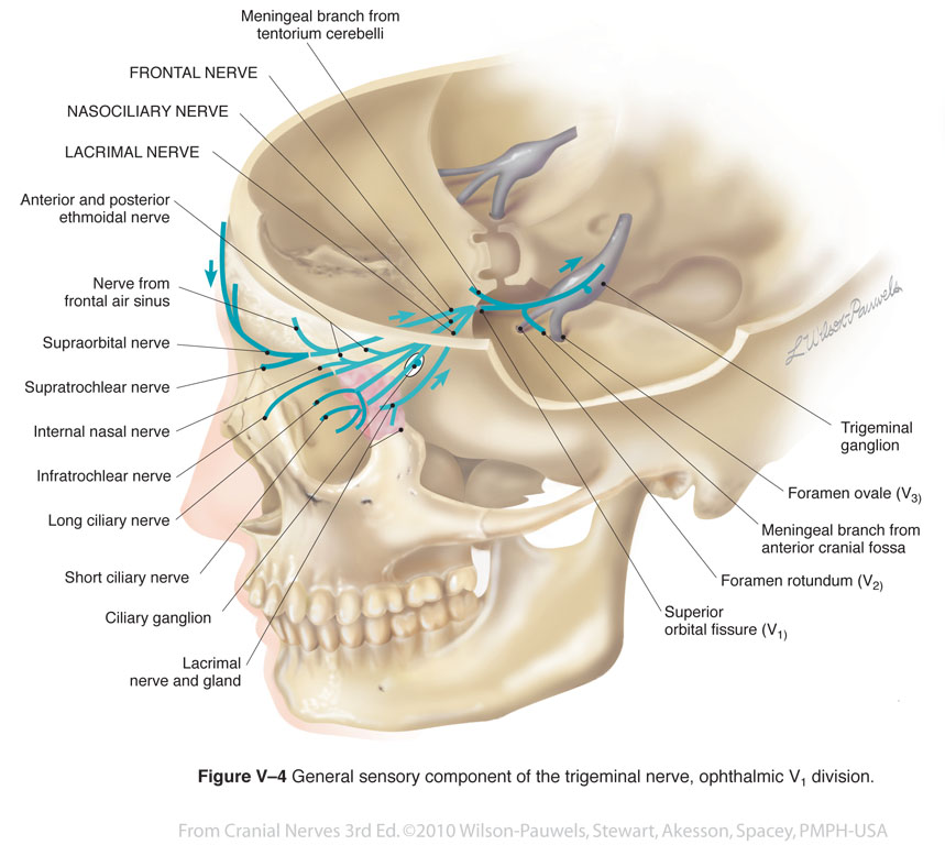
| Figure V-4 General sensory component of the trigeminal nerve, ophthalmic (V1) division. General sensory afferent (blue-green) |
 |
 |
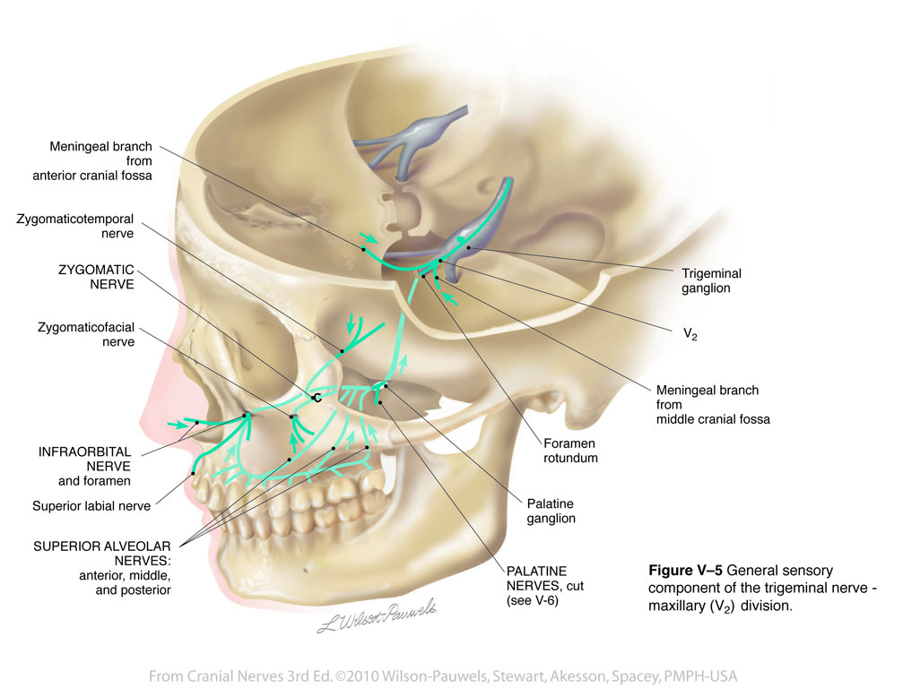
| Figure V-5 General sensory component of the trigeminal nerve, maxillary (V2) division. General sensory afferent (blue-green) |
 |
 |
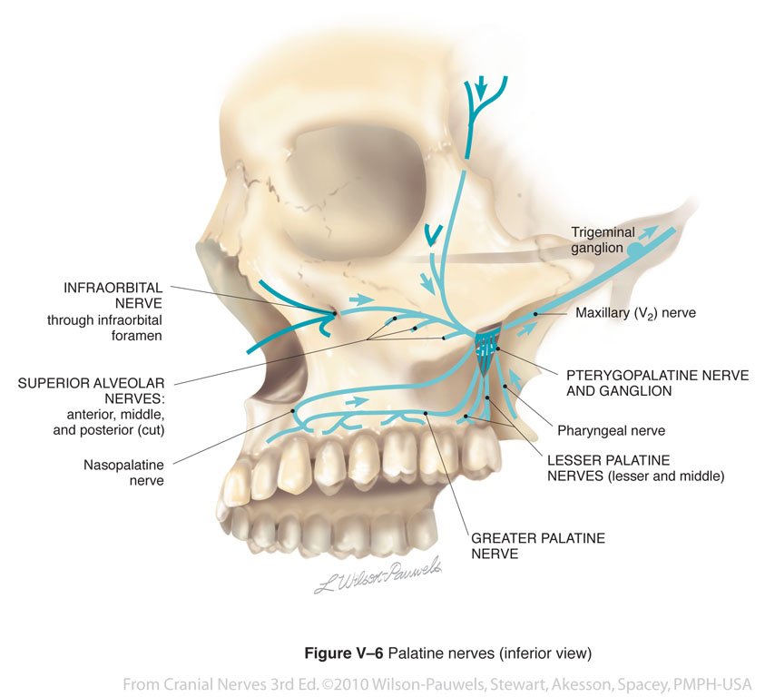
| Figure V-6 Palatine nerves (inferior view). General sensory afferent (blue-green) |
 |
 |
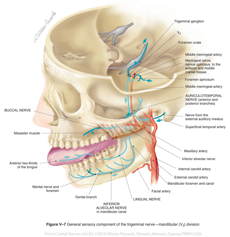
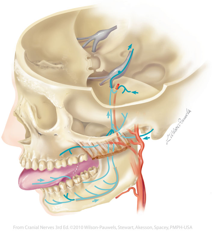
| Figure V-7 General sensory component of the trigeminal nerve - mandibular (V3) division. General sensory afferent (blue-green) |
 |
 |
| Figure V-8 Trigeminal sensory nucleus (dorsal view of the brain stem). | 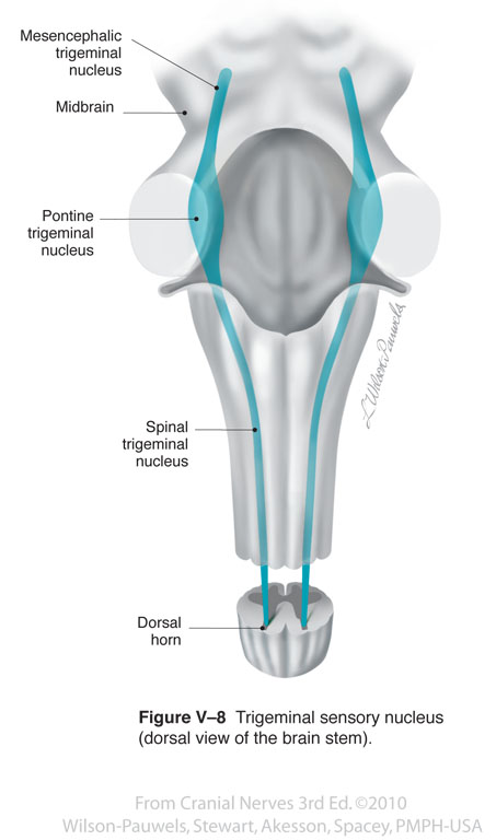 |
 |
| Figure V-9 Trigeminal sensory nucleus (lateral view of the brain stem). | 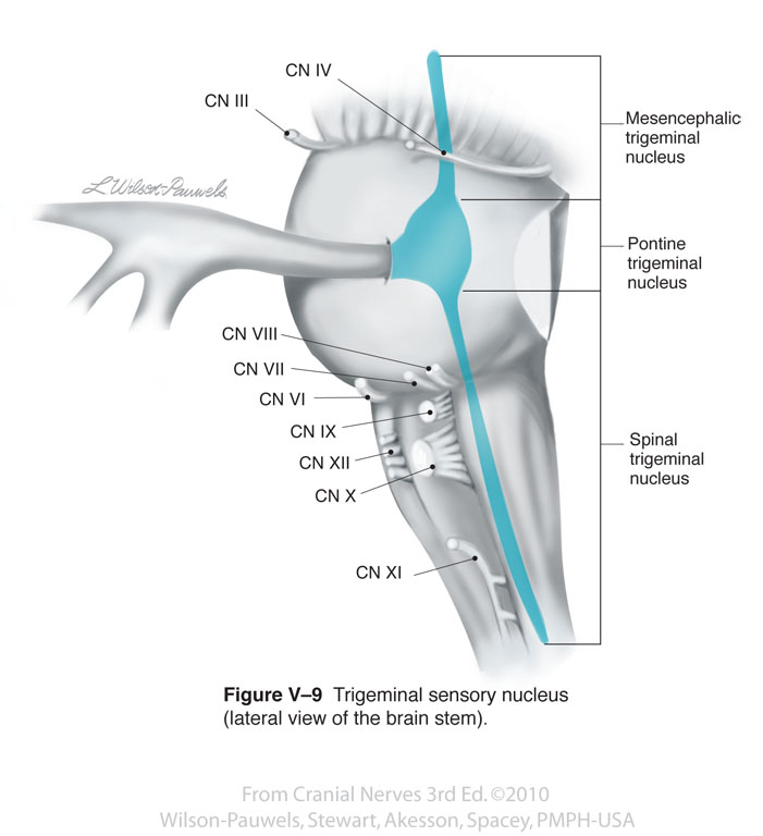 |
 |
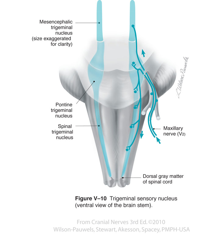
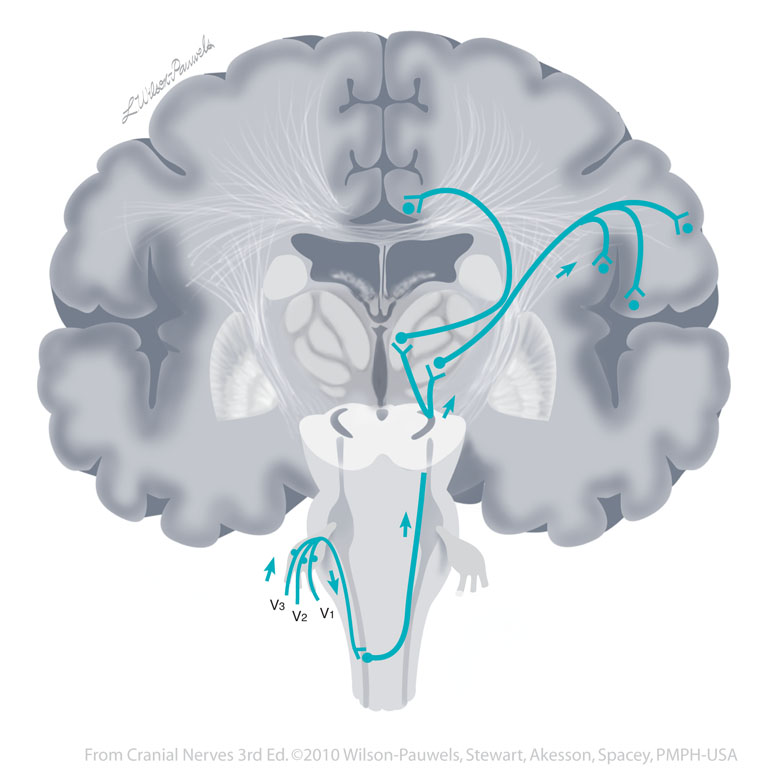
| Figure V–10 Trigeminal sensory nucleus (ventral view of the brain stem). General sensory afferent (blue-green) |
 |
 |
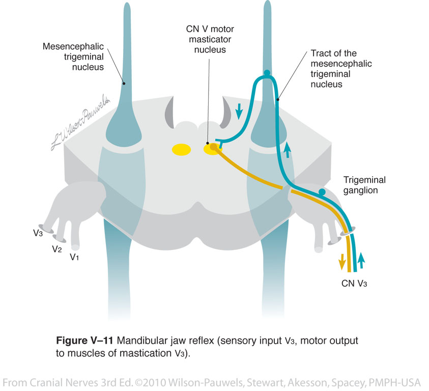
| Figure V-11 Mandibular jaw reflex (sensory input V3, motor output to muscles of mastication V3). General sensory afferent (blue-green) Branchial motor efferent (yellow) |
 |
 |
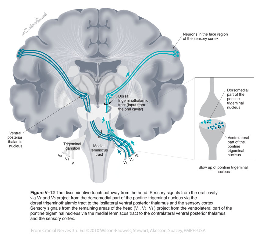
| Figure V–12 The discriminative touch pathway from the head. Sensory signals from the oral cavity via V2 and V3 project from the dorsomedial part of the pontine trigeminal nucleus via the dorsal trigeminothalamic tract to the ipsilateral ventral posterior thalamus and the sensory cortex. Sensory signals from the remaining areas of the head (V1, V2, V3 ) project from the ventrolateral part of the pontine trigeminal nucleus via the medial lemniscus tract to the contralateral ventral posterior thalamus and the sensory cortex. General sensory afferent (blue-green) |
 |
 |
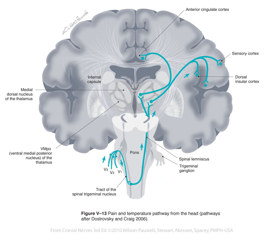
| Figure V-13 Pain and temperature pathways from the head (pathways after Dostrovsky and Craig 2006). General sensory afferent (blue-green) |
 |
 |
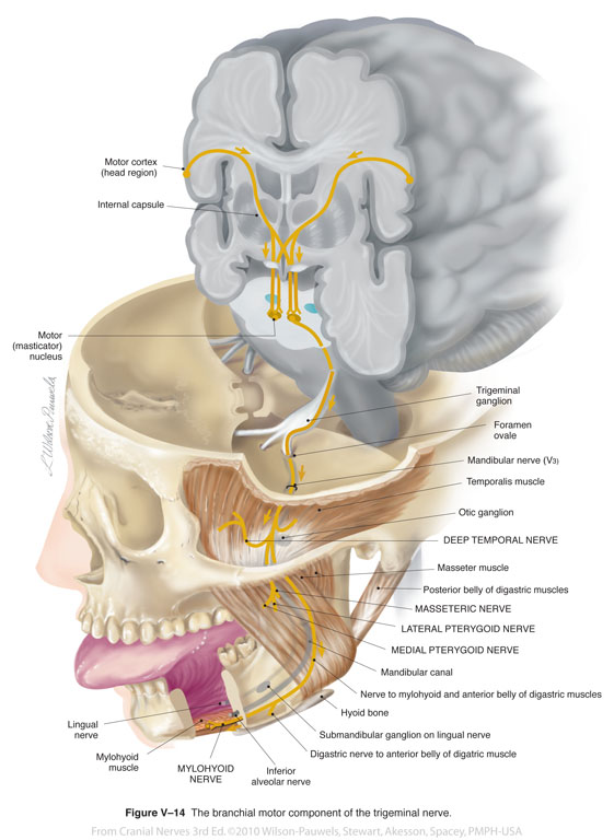
| Figure V-14 The branchial motor component of the trigeminal nerve. Branchial motor efferent (yellow) |
 |
 |
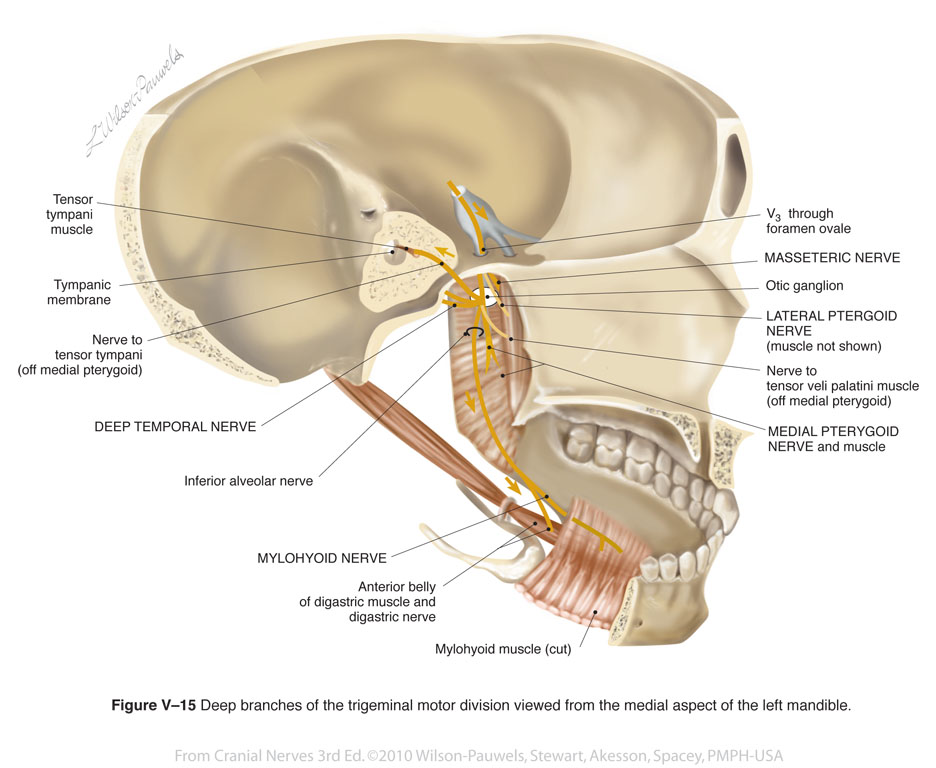
| Figure V-15 Deep branches of the trigeminal motor division viewed from the medial aspect of the left mandible. Branchial motor efferent (yellow) |
 |
 |
| Figure V–16 Mary’s MRI showed a meningioma growing on her left petrous temporal ridge. The tumor is pressing on her pons at the entry of the trigeminal nerve. A, MRI scan. B, The tumor has been artifically coloured (red) and representations of the positions of the trigeminal nerves have been ghosted in to show that the left trigeminal nerve (blue) would be compressed by the meningioma. (Image courtesy of Dr. David Mikulis.). | 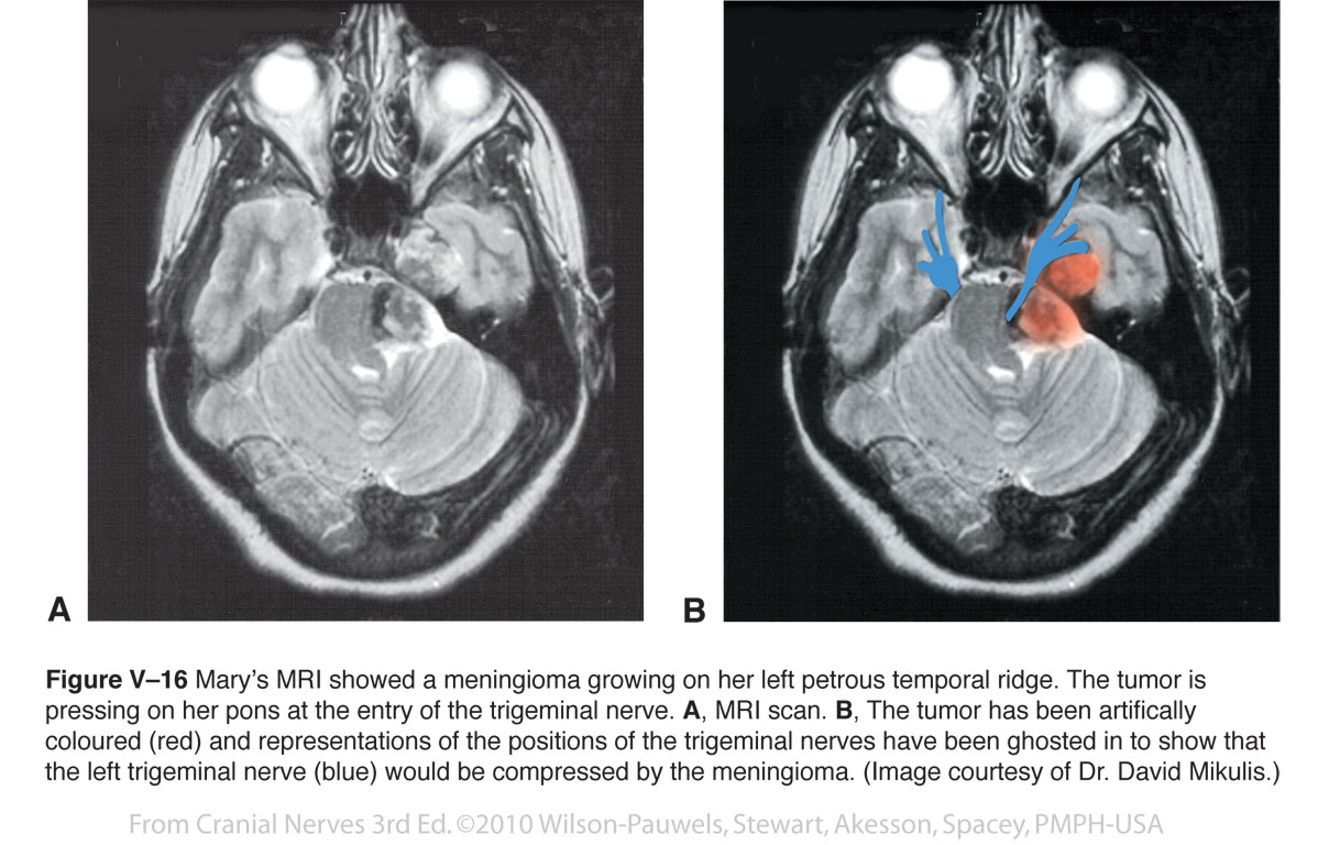 |
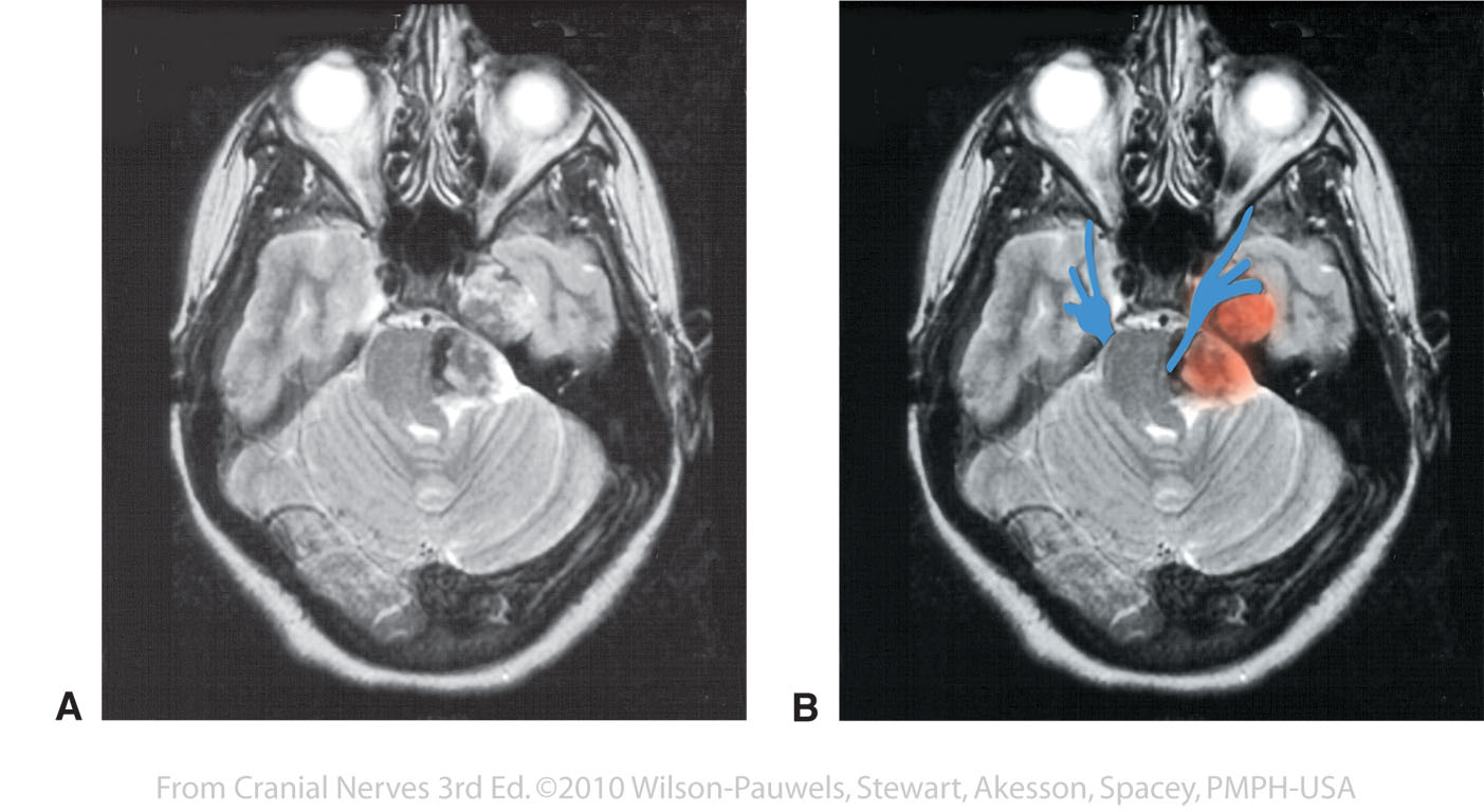 |
| Figure V-17 Schwannoma: an enlarged tumor in the cerebellopontine angle compressing the root of the trigeminal nerve (sagittal section through the jugular foramen). General sensory afferent (blue-green) Branchial motor efferent (yellow) |
 |
 |
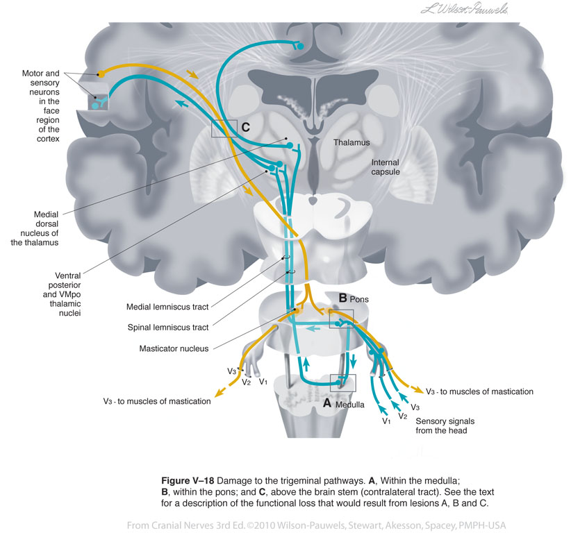
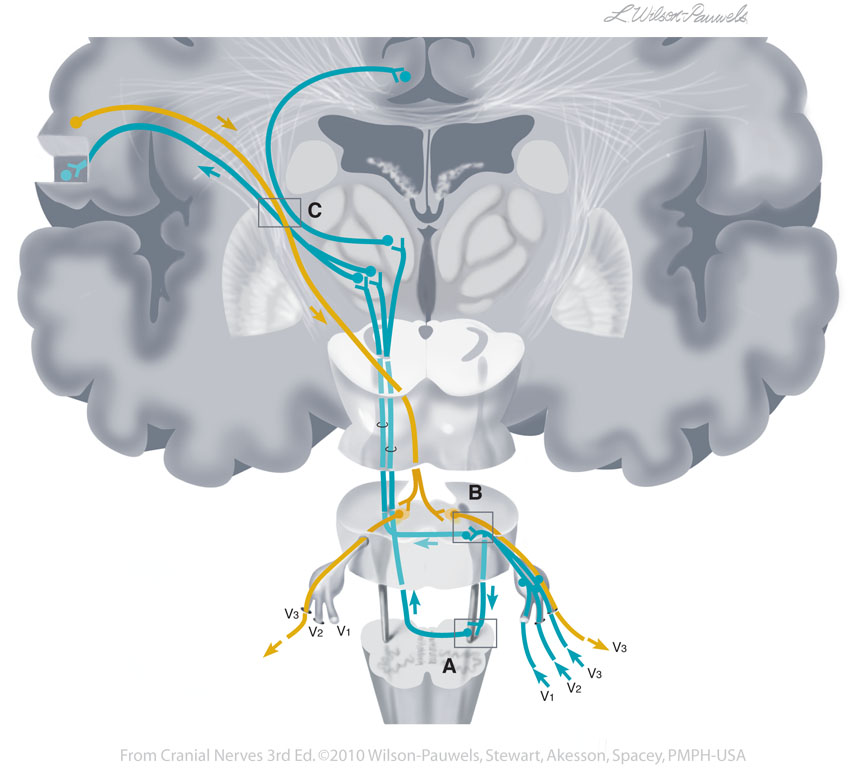
| Figure V-18 Damage to the trigeminal pathways. A, within the medulla; B, within the pons; and C, above the brainstem (contralateral tract). General sensory afferent (blue-green) Branchial motor efferent (yellow) |
 |
 |
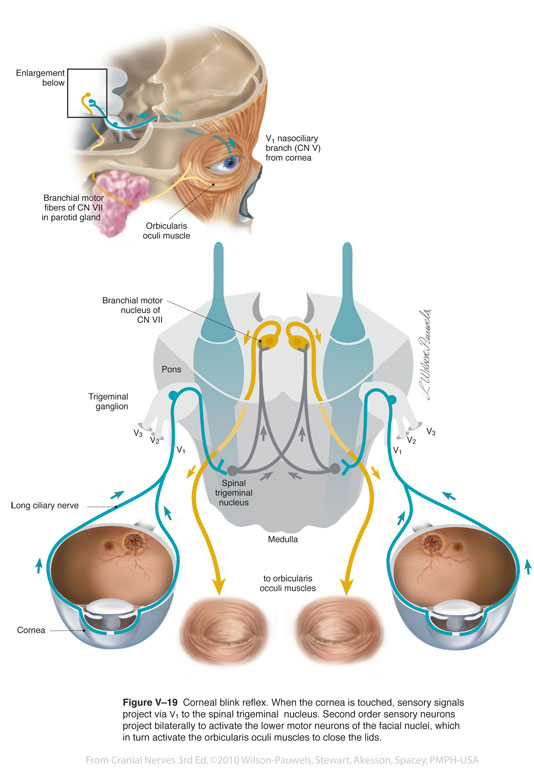
| Figure V–19 Corneal blink reflex. When the cornea is touched, sensory signals project via V1 to the spinal trigeminal nucleus. Second order sensory neurons project bilaterally to activate the lower motor neurons of the facial nuclei, which in turn activate the orbicularis oculi muscles to close the lids. General sensory afferent (blue-green) Branchial motor efferent (yellow) |
 |
 |
| Figure V-20 Clinical testing for sensation. General sensory afferent (blue-green) |
 |
 |
| Figure V-21 Testing the corneal reflex. | 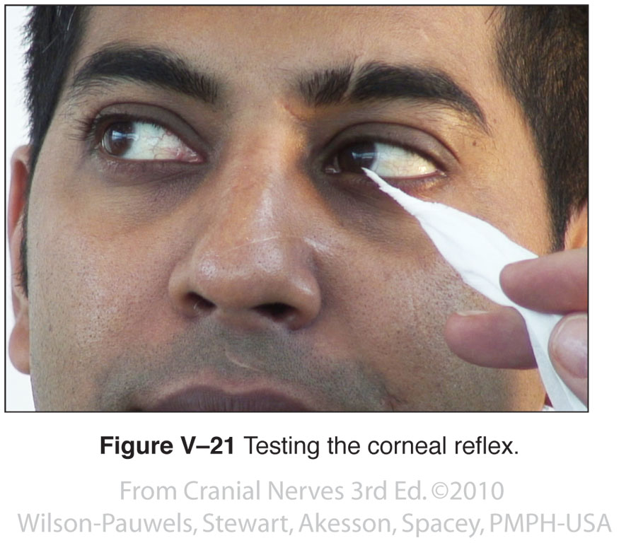 |
 |
| Figure V-22 Clinical testing for muscles of mastication. | 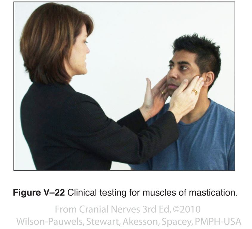 |
 |