Vestibulocochlear VIII
| Description | Labelled | Unlabelled |
|---|---|---|
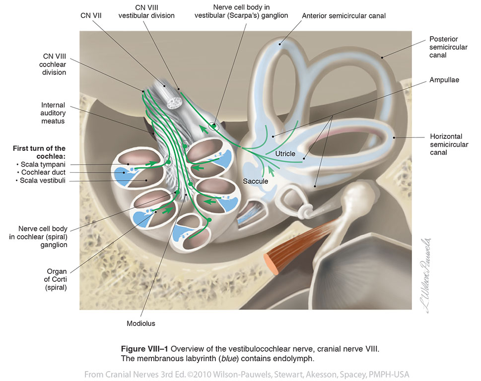
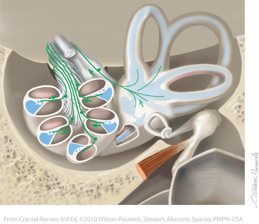
| Figure VIII–1 Overview of the vestibulocochlear nerve, cranial nerve VIII. The membranous labyrinth (blue) contains endolymph. Special sensory afferent (green) |
 |
 |
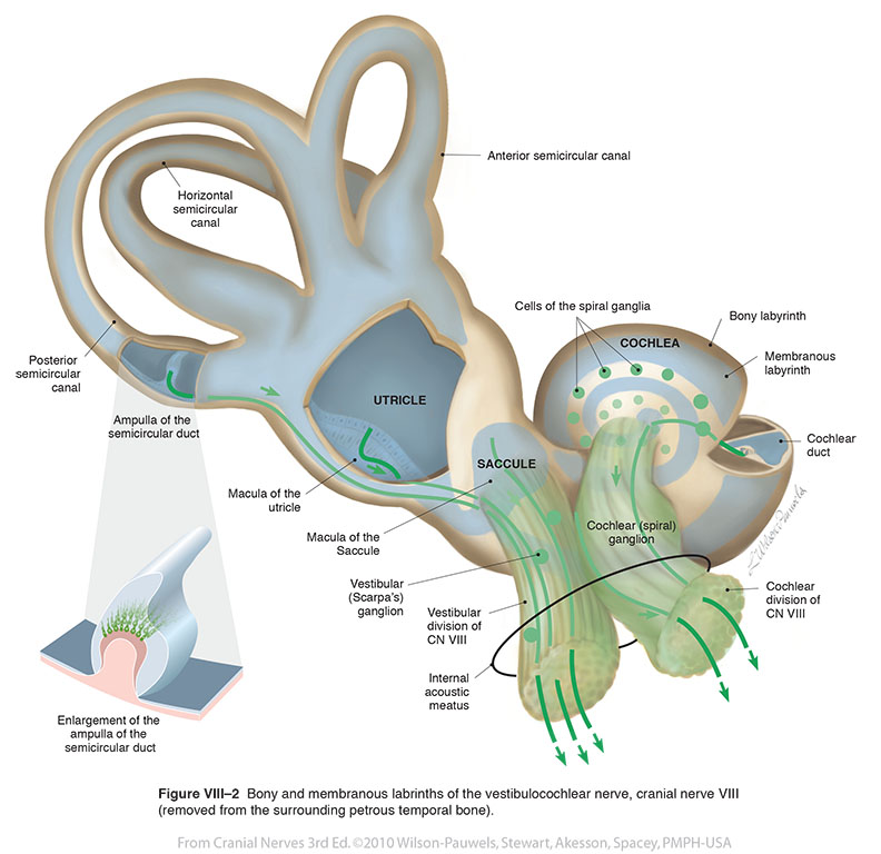
| Figure VIII–2 Bony and membranous labyrinths of the vestibulocochlear nerve, cranial nerve VIII (removed from the surrounding petrous temporal bone). Special sensory afferent (green) |
 |
 |
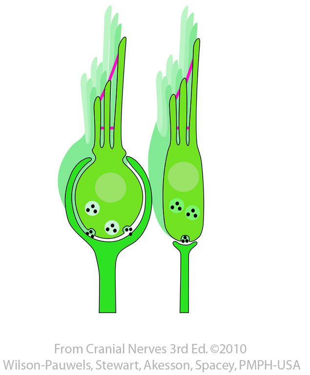
| Figure VIII–3 Type 1 and type 2 hair cells in vestibular apparatus. | 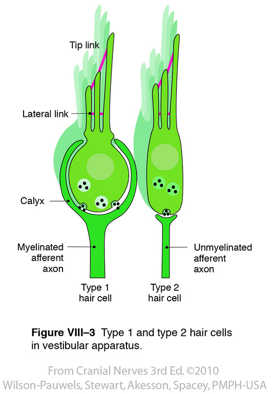 |
 |
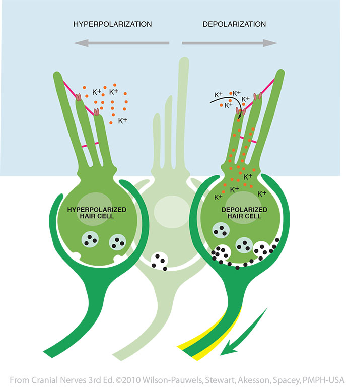
| Figure VIII–4 Sensory transduction in hair cells. | 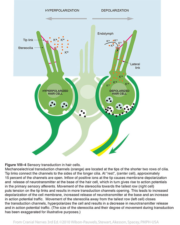 |
 |
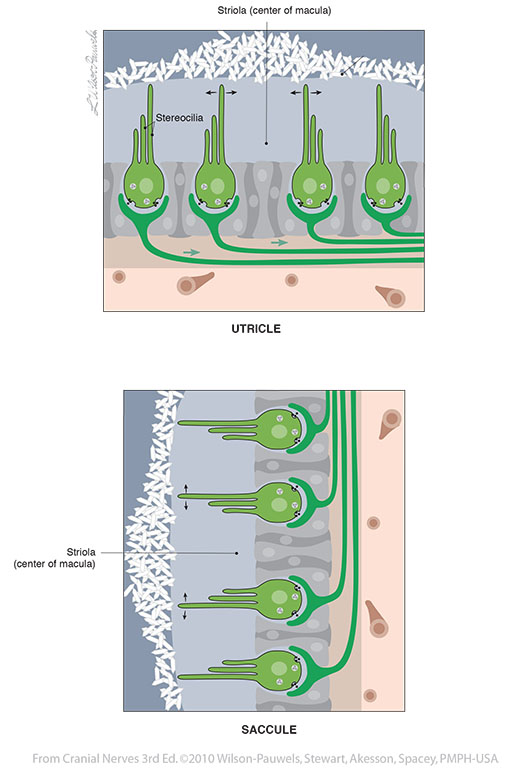
| Figure VIII–5 Macular hair cells of the utricle and saccule. | 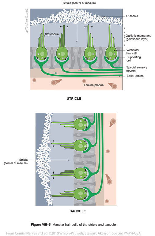 |
 |
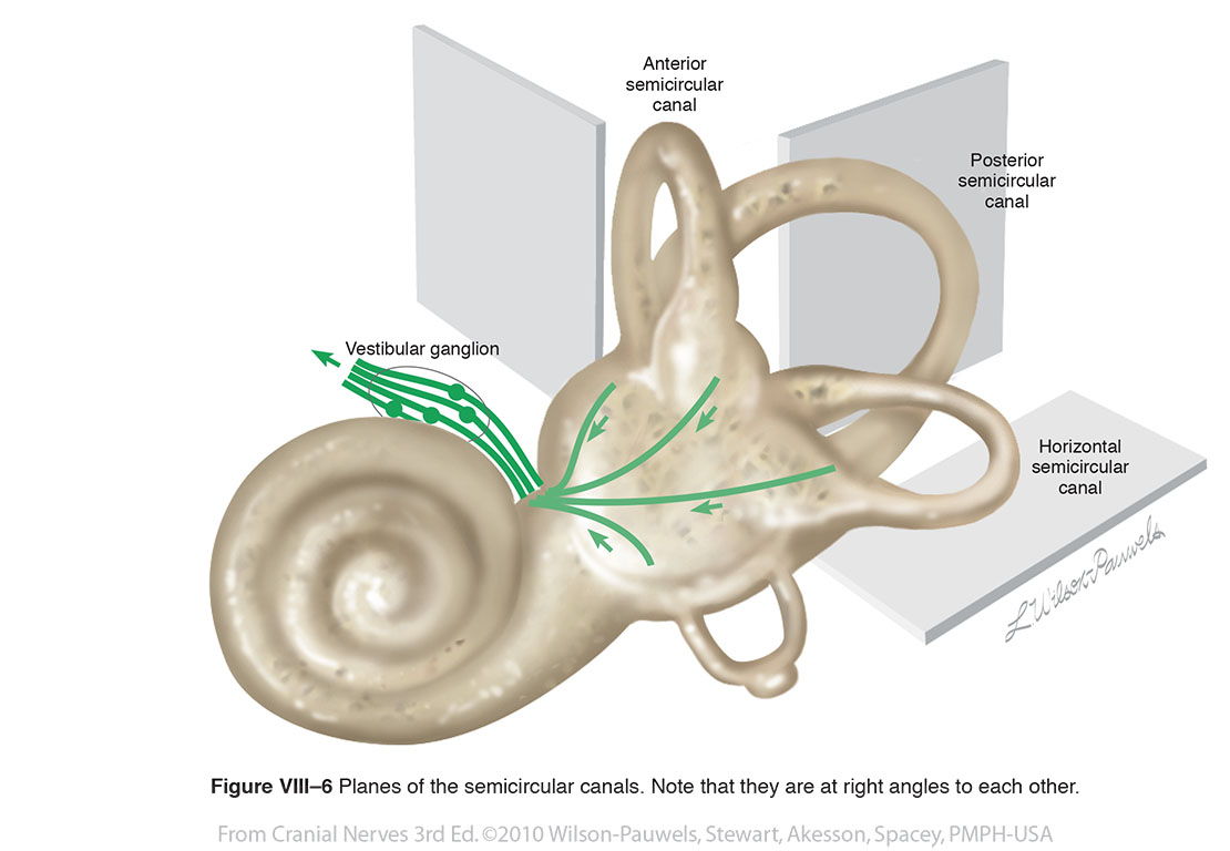
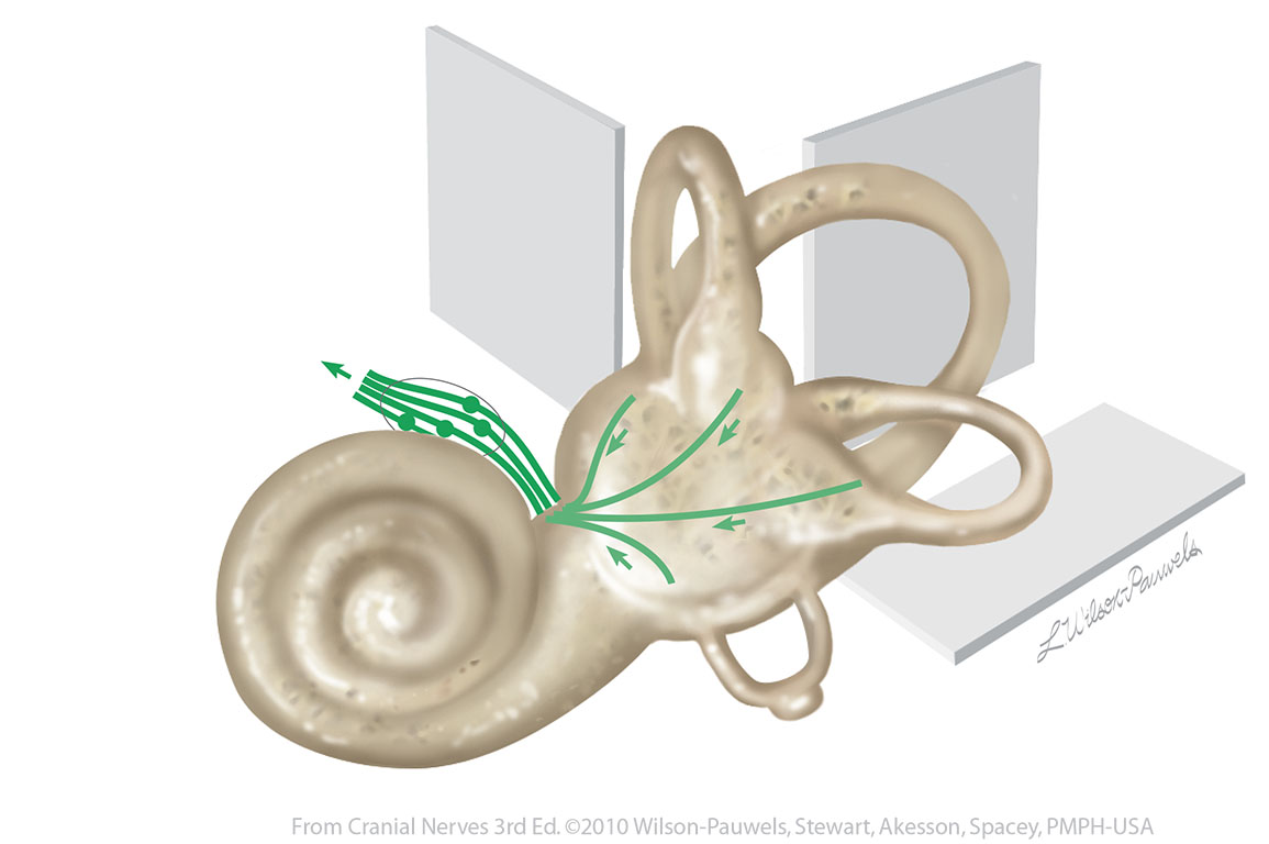
| Figure VIII–6 Planes of the semicircular canals. Note that they are at right angles to each other. Special sensory afferent (green) |
 |
 |
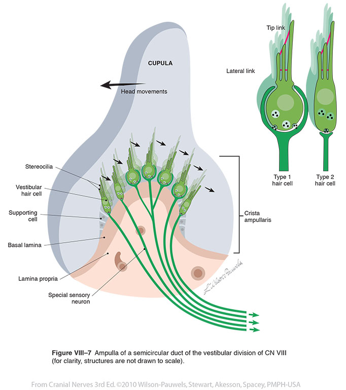
| Figure VIII–7 Ampulla of a semicircular duct of the vestibular division of CN VIII (for clarity, structures are not drawn to scale). Special sensory afferent (green) |
 |
 |
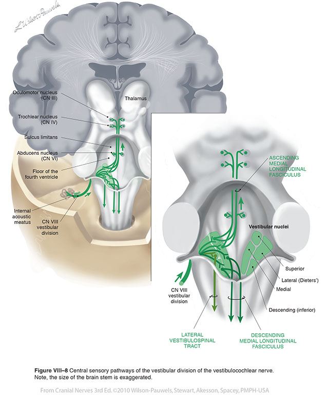
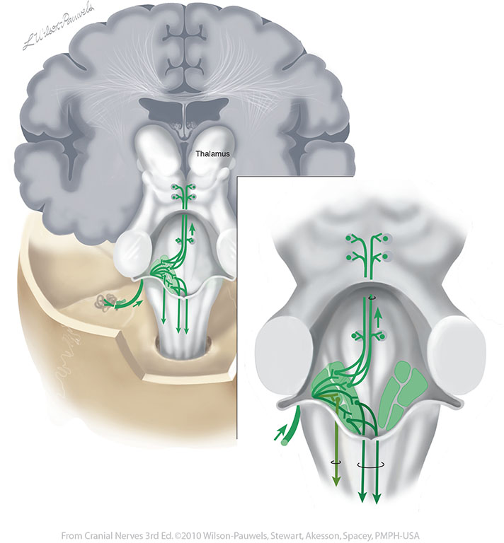
| Figure VIII–8 Central sensory pathways of the vestibular division of the vestibulocochlear nerve. Note, the size of the brain stem is exaggerated. Special sensory afferent (green) |
 |
 |
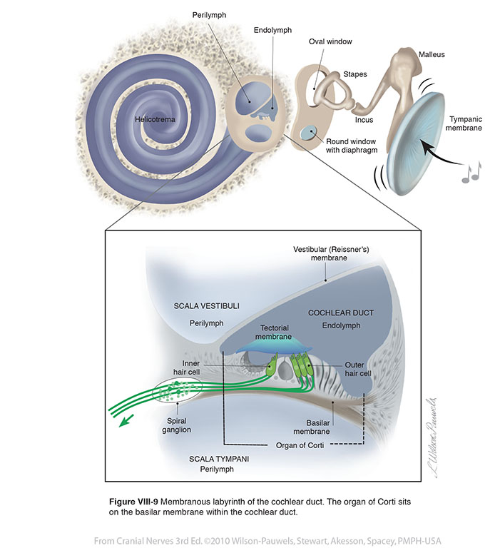
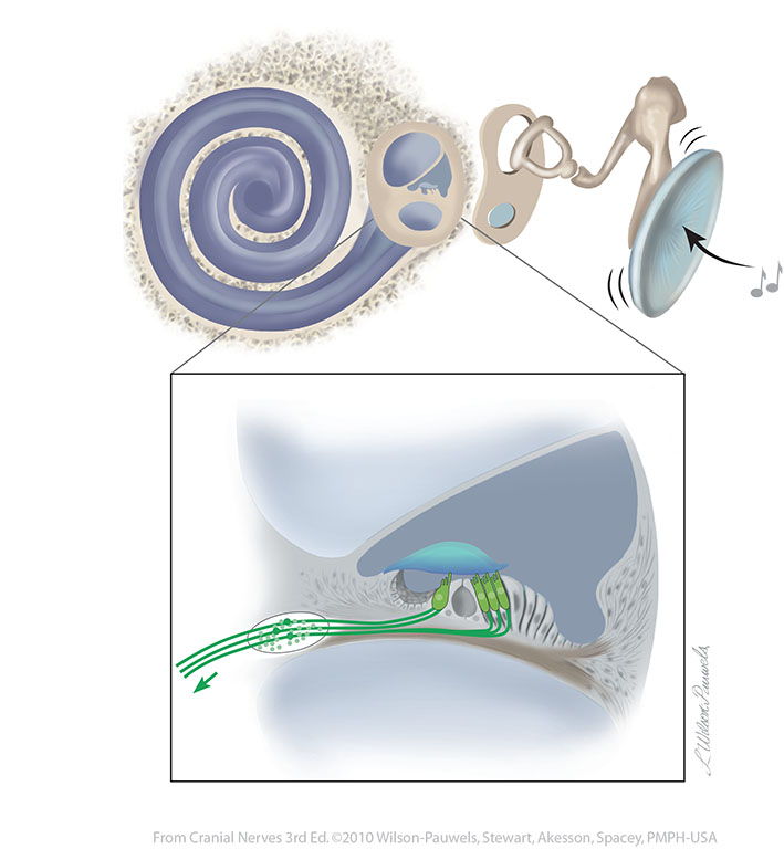
| Figure VIII-9 Membranous labyrinth of the cochlear duct. The organ of Corti sits on the basilar membrane within the cochlear duct. Special sensory afferent (green) |
 |
 |
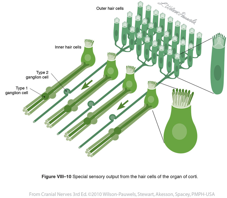
| Figure VIII–10 Special sensory output from the hair cells of the organ of corti. Special sensory afferent (green) |
 |
 |
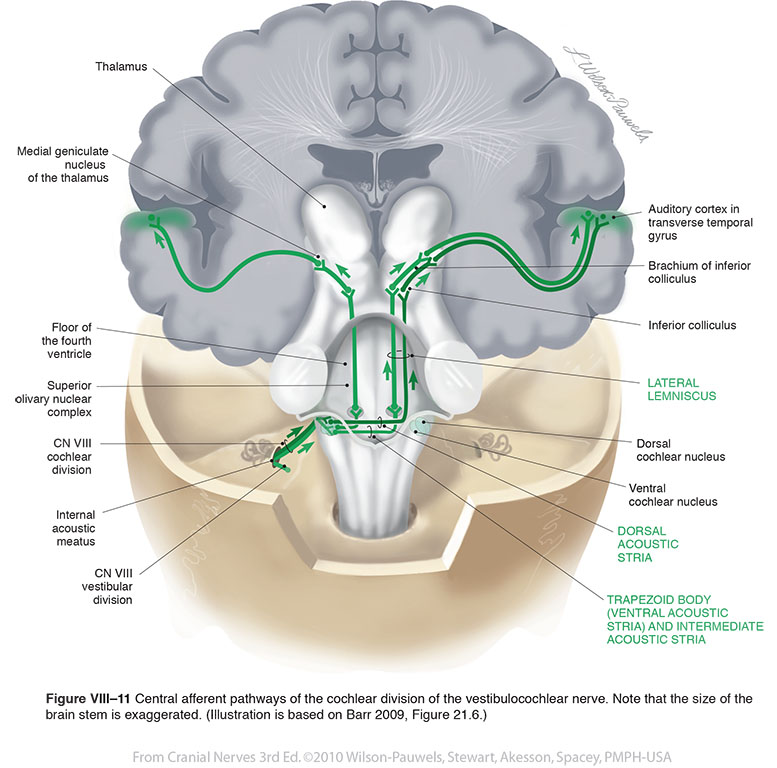
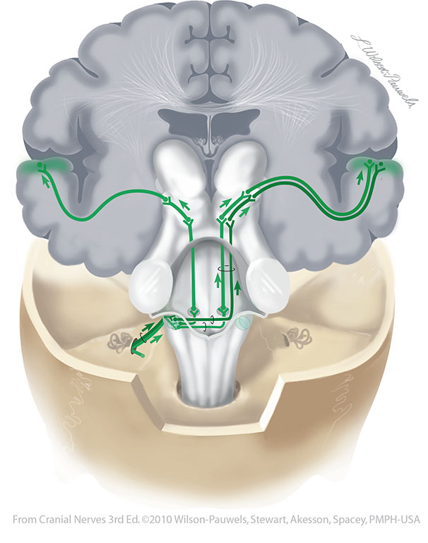
| Figure VIII–11 Central afferent pathways of the cochlear division of the vestibulocochlear nerve. Note that the size of the brain stem is exaggerated. (Illustration is based on Barr 2009, Figure 21.6.). Special sensory afferent (green) |
 |
 |
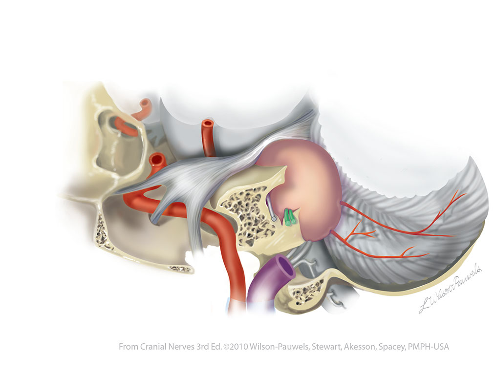
| Figure VIII–12 An enlarged tumor (schwannoma) in the cerebellopontine angle compromising cranial nerves V, VII, and the vestibular and cochlear divisions of VIII (illustrated in green). This is a sagittal section through the jugular foramen. | 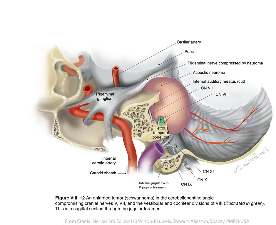 |
 |
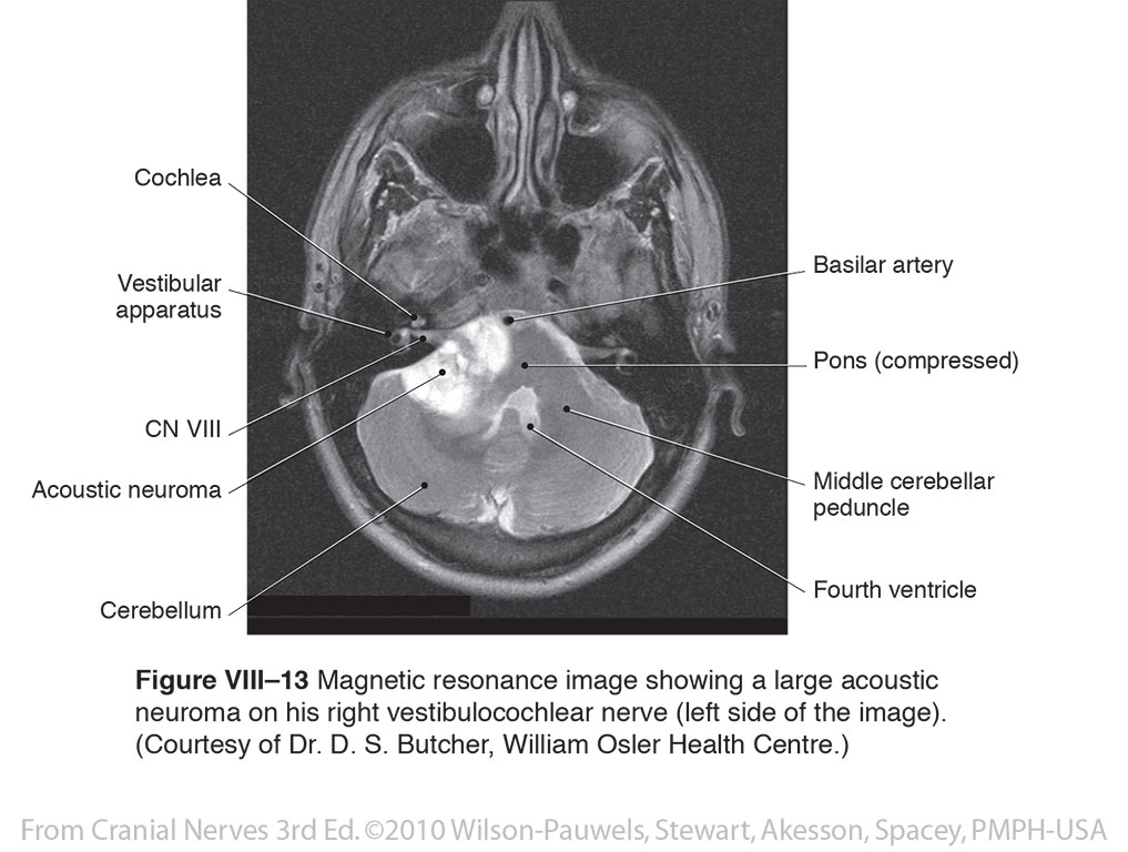
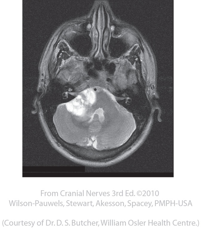
| Figure VIII–13 Magnetic resonance image showing a large acoustic neuroma on his right vestibulocochlear nerve (left side of the image). (Courtesy of Dr. D. S. Butcher, William Osler Health Centre.) |
 |
 |
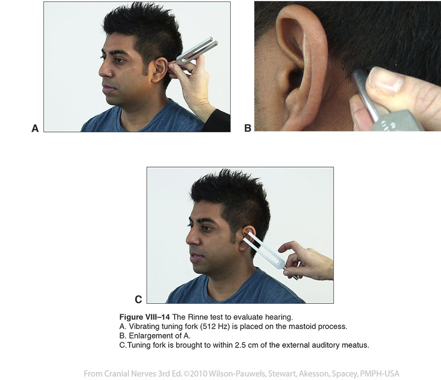
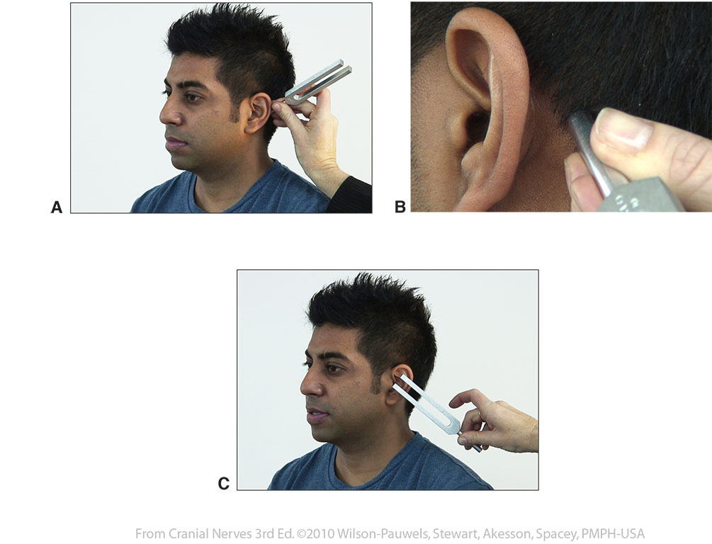
| Figure VIII–14 The Rinne test to evaluate hearing. A. Vibrating tuning fork (512 Hz) is placed on the mastoid process. B. Enlargement of A. C.Tuning fork is brought to within 2.5 cm of the external auditory meatus. |
 |
 |
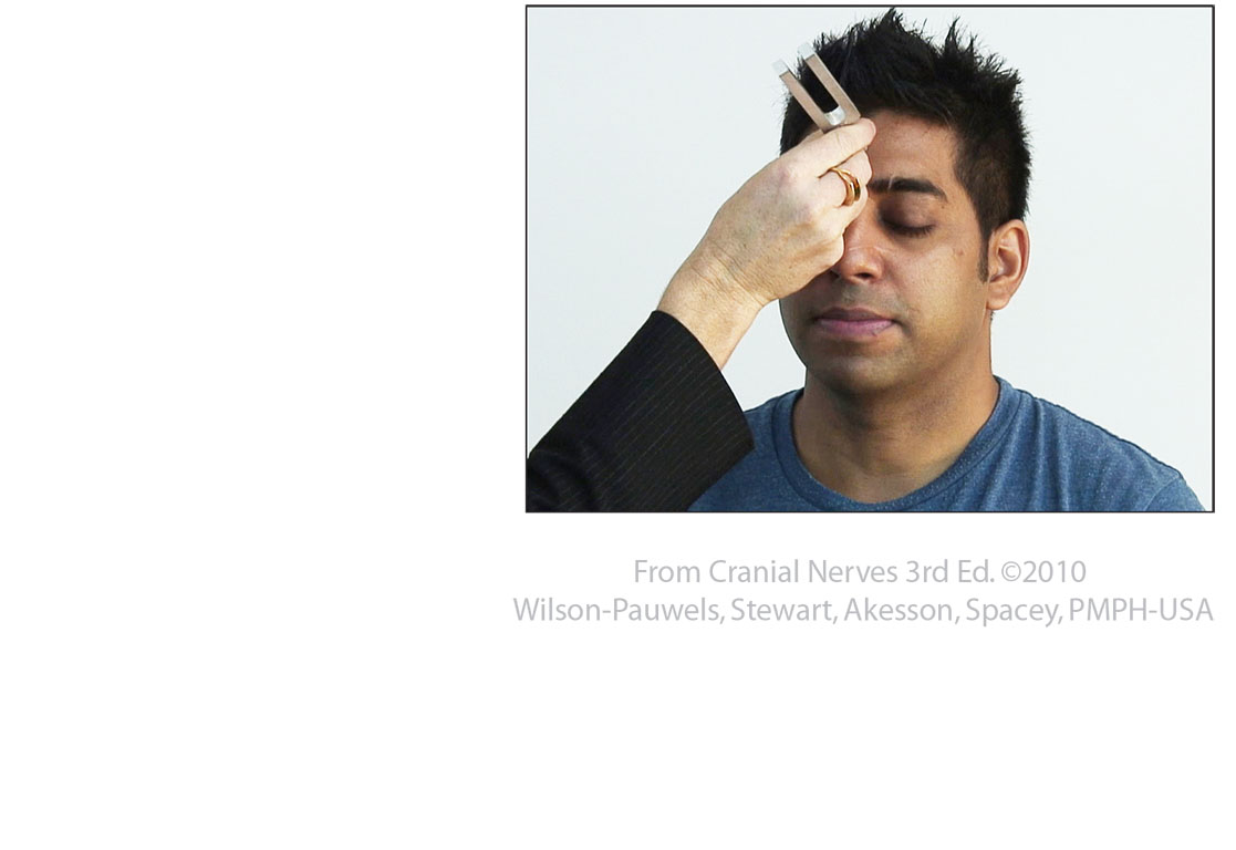
| Figure VIII–15 Weber’s test to distinguish between conductive and sensorineural deafness. A vibrating tuning fork (512 Hz) is placed in the middle of the forehead. | 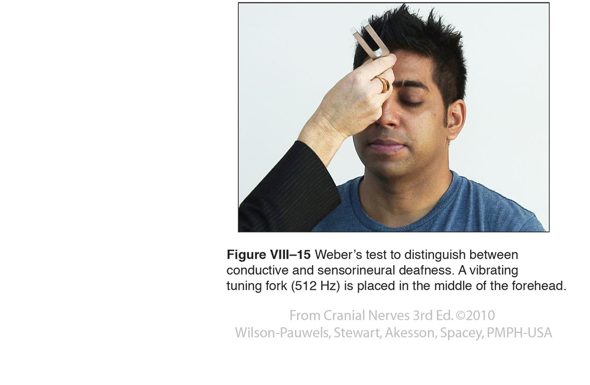 |
 |